Publication Date: November 5, 2026
Access counts:0
Next-Generation Imaging Using Pulsed Neutrons
-Structure-Resolved Imaging Enabled by Neutron Radiography and Resonance Analysis-
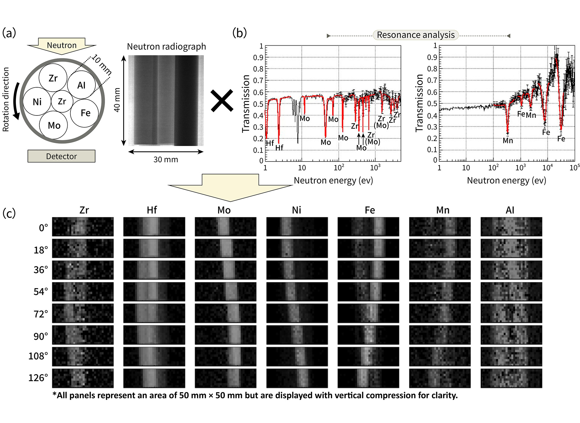
Fig. 1 Neutron radiographs of six metal rods (a) and resonance measurements (b), along with the results of resonance imaging
—which combines these two techniques—to identify the individual metal rods in the rotating sample (c)
At the J-PARC MLF, which provides users with high-intensity neutron beams, a wide variety of research projects that utilize the unique properties of neutrons are carried out daily. One of the characteristics of the MLF, one of the world’s leading neutron facilities, is that neutrons with energies ranging from the keV to the MeV are readily available.
Neutron resonance reactions are characteristic of this energy range. Neutron resonance absorption by atomic nuclei exhibits a resonance absorption spectrum characteristic of each nuclide; therefore, by analyzing resonance peaks and other spectral features, it is possible to determine which elements are present in a sample and in what quantities.
Resonance imaging, which combines this technique with two-dimensional neutron transmission imaging, enables the visualization of the spatial distribution of elements within a sample and their respective amounts (Fig. 1). In recent years at the MLF, measurement efficiency has improved through the expansion of the analyzed energy range, increased sensitivity to a wider range of elements, and the introduction of methods that analyze not only resonance peaks but also the entire measured energy spectrum. Consequently, shorter resonance imaging times are becoming feasible.
Neutrons are particularly effective for visualizing the internal structures of dense materials, such as metals, that are difficult for X-rays to penetrate. Taking advantage of this characteristic, resonance imaging has been applied to fields ranging from the elemental mapping and quantitative analysis of simulated molten debris relevant to the decommissioning of the Fukushima Daiichi Nuclear Power Station to engineering research and industrial applications.
Recently, it has also begun to be used in the study of artifacts and fossils, and it is expected to find applications even more broadly across various fields.

Comments on This Article
Share your comments or feedback on this article using the comment form.