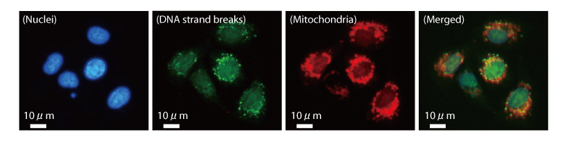
Fig.4-15 DNA strand breaks in mitochondria of mammalian cells following γ ray irradiation
Radiation-induced DNA strand breaks in mitochondria of mammalian cells were visualized by employing PprA protein using an immunofluorescence technique. Colocalization of fluorescence breaks and mitochondria was observed in the image merging the two (right panel). This image suggests that DNA strand breaks existing in mitochondria were effectively detected by utilizing DNA strand break binding ability of PprA protein.
Go back by your web browser, or click the right button. « Close