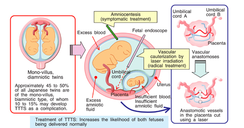
Fig.11-7 Example of congenital disease: Twin-to-Twin Transfusion Syndrome (TTTS)
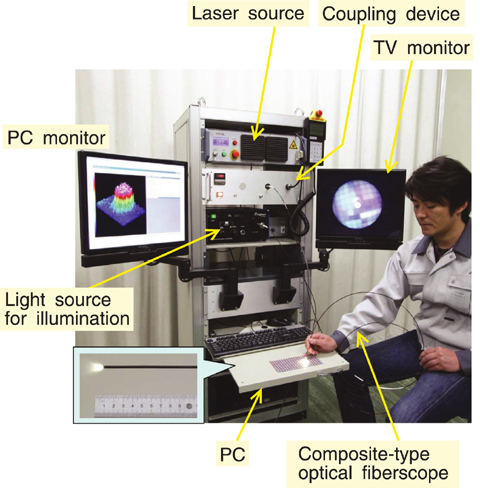
Fig.11-8 Extra-fine endoscope having a laser radiation function
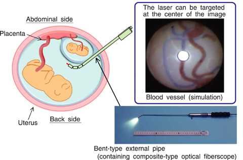
Fig.11-9 Technology that enables placenta treatment at all positions
Twin to twin transfusion syndrome (popular name TTTS) is one of the congenital diseases that twins develop in utero. This disease that only occurs in cases consisting of two amnions and one placenta (mono-villus diamniotic twins) requires highly advanced medical treatment. In the case of normal mono-villus diamniotic-twins, each blood vessel is connected to the placenta and the balance of blood circulating through each is good. When blood flow between twins becomes unbalanced, and blood continues flowing to only one fetus, TTTS develops. A laser treatment method while observing with an endoscope (popular name FLP) recently is attracting attention as a radical therapy of TTTS, (Fig.11-7).
However, experience in FLP treatment is superficial nationwide, and the reality is that there are only a few hospitals nationwide that can perform FLP treatment. In addition, there are the following problems because this treatment is intended for a fetus floating in the uterus (in amniotic fluid) which is a small space; (1) the definite vascular configuration (a map) of connection to the placenta is not clear, (2) the distance between the tip of the optical fiber and a blood vessel is not clear, (3) quantitative determination whether the bloodstream is stopped or not is not possible, (4) the endoscope image is narrow, and (5) it is hard if the placenta is in front of the fetuses (placenta previa). Advanced technical progress and a highly skilled doctor are necessary to do such an operation with the conventional endoscope.
Therefore to make a contribution to FLP treatment, by utilizing a composite-type optical fiber by which observation and laser irradiation are possible in one optical fiber at the same time, we produced a device which promises to solve the problems with the conventional endoscope (Fig.11-8). We note that this device was created as a spin-off from development of a robot for inspecting and maintaining nuclear fusion reactors.
In this system, the middle of an image can be laser-irradiated, and there are functions to measure the distance to an object and blood quantity/flow speed. This allows laser radiation to be applied precisely, safely and comparatively easily. This instrument has potential for prenatal surgical treatment of the congenital disease mentioned above. In addition, this makes laser radiation easy even in the case of placenta previa (Fig.11-9). Furthermore, because the diameter of the hole (possibly through the abdomen) can be reduced by reducing the endoscopic external diameter to as little as 2 mm, we can expect that the burden to a mother's body will be reduced greatly (Minimally Invasive Treatment). The efficacy of this system has already been confirmed in animal experiments. We are proceeding with development for clinical applications now.
<Previous: 11-1 | Next: 12 Computational Science and E-Systems Research >

