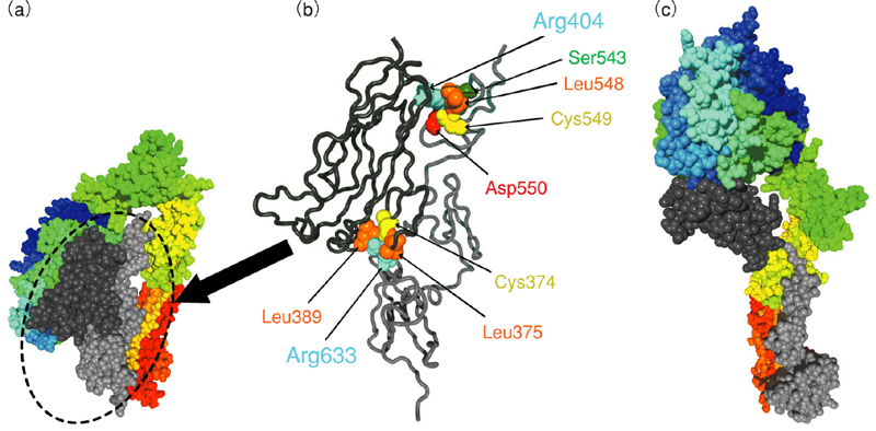
Fig.12-6 (a) and (b) X-ray crystal structure of integrin αVβ3; (c) computer model of the extended conformation
(a) 3D structure of integrin solved by X-ray crystal structure analysis: ball model of the molecule when "bowing down".
(b) A Magnified tube model of part of the integrin structure. Important amino-acid residues are shown by ball model and their names are shown.
(c) Extended conformation of integrin obtained by computer simulation of large-scale conformational change.
(b) A Magnified tube model of part of the integrin structure. Important amino-acid residues are shown by ball model and their names are shown.
(c) Extended conformation of integrin obtained by computer simulation of large-scale conformational change.
Go back by your web browser, or click the right button. « Close