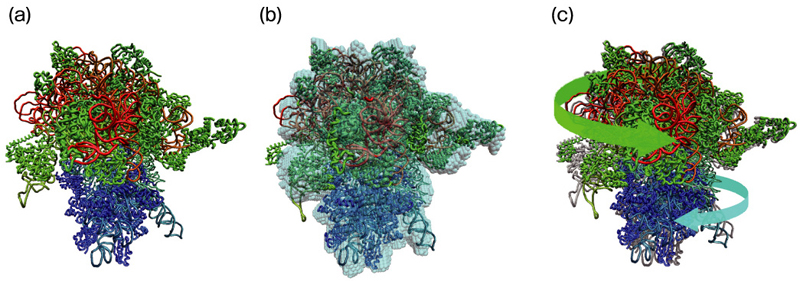
Fig.4-24 Three-dimensional structures of a ribosome determined by different methods
(a) X-ray crystal structure of the ribosome. It is composed of large (red, green) and small (blue) subunits.
(b) 3D electron microscopy structure (cyan), shown overlaid upon a best-fit molecular model.
(c) Comparison of (a) the X-ray crystal structure (gray) with (b) the best-fit molecular model. The latter is twisted along the direction of the arrows with respect to the former.
Go back by your web browser, or click the right button.« Close