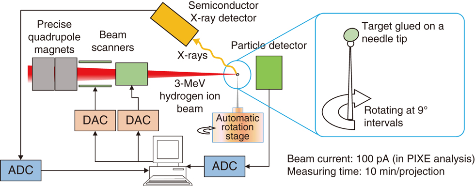
Fig.5-25 Schematic diagram of the PIXE Tomography system
A 3-MeV hydrogen-ion beam is focused to a diameter of 1 μm by precise quadrupole magnets and used to scan across a 100 μm × 100 μm area on a target. The three-dimensional elemental distribution is reconstructed from the location and energy of the X-rays emitted from the target by a computer.
Go back by your web browser, or click the right button.« Close