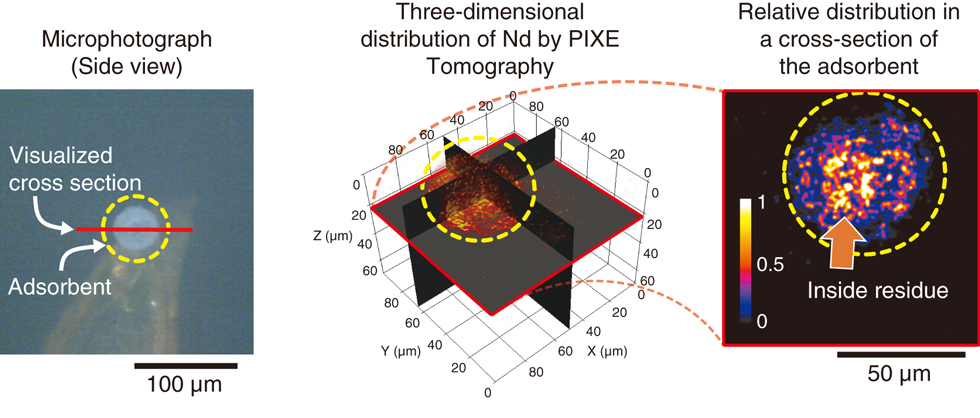
Fig.5-26 Optical micrograph of a porous silica-adsorbent particle for EXC (left figure) and the results of PIXE tomography (center and right figures)
The left figure exhibits a porous silica-adsorbent particle fixed on the tip of a needle. An arbitrary tomographic image of the Nd distribution in the particle was obtained by PIXE tomography, as shown in the right figure.
Go back by your web browser, or click the right button.« Close