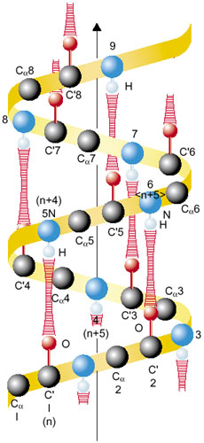
According to Pauling's model, the alpha-helix structure of proteins is often illustrated in textbooks by the formation of straight N-H---O hydrogen bonds between the N-H group in amino acid residue n+4 and the C-O group in residue n (Fig. 4-13). However, this picture is oversimplified. While recent high resolution X-ray crystallographic studies offer some positional information on the hydrogen atoms, we do not yet have a satisfactory understanding of hydrogen bonds in the alpha-helices of proteins, because the positional data for hydrogen atoms in alpha-helices are not yet complete.
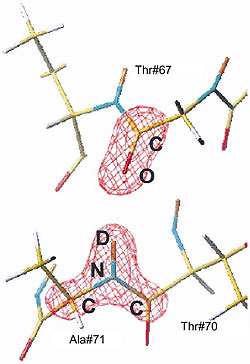
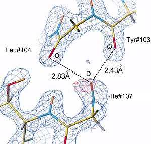
On the other hand it is well known that neutron diffraction provides an experimental method for directly locating hydrogen atoms. Recently, we have collected high resolution neutron diffraction data from tetragonal HEWL (hen egg-white lysozyme) and myoglobin, and the positions of all the hydrogen atoms in both of the proteins have been determined accurately. All the hydrogen bonds between C=O and N-H in the helices were surveyed, including the location of the hydrogen atoms. Figure 4-15 shows one example of how a conventional hydrogen bond in an alpha-helix of myoglobin is seen in a neutron diffraction experiment. Moreover, we found several bifurcated hydrogen bonds, where there is a formation of hydrogen bonds between the N-H group in amino acid residue n+4 and not only the C-O group in residue n, but also that in n+1. A typical example of a bifucated hydrogen bond in myoglobin is illustrated in Fig. 4-15.
The existence of bifurcated hydrogen bonds clearly indicates that further accumulation of results from high-resolution neutron protein crystallography will provide a more complete understanding of the nature of hydrogen bonds in proteins in the future. |