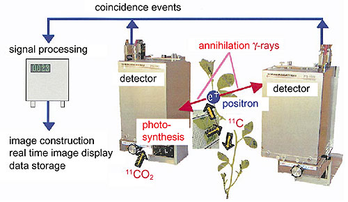
Sensing environmental change, plants can control their metabolism and adapt to the change. Sometimes plants modify their form to survive a new environmental condition. To understand their survival strategies, a new measurement technique has been developed which allows us to observe the translocation of materials in a living plant subjected to different conditions. Applying the same principle as that of positron emission tomography (PET) in medicine, which analyses gamma-rays emitted during the annihilation of positrons and is used for diagnosis of cancers and human brain functions, we have constructed a positron emitting tracer imaging system (PETIS) as shown in Fig. 9-10. The system is able to map a two-dimensional distribution of compounds labeled with a positron-emitting tracer in a plant as a real time image with a spatial resolution of 2.3 mm. The signal-to-noise ratio of PETIS is higher than that of PET, because gamma-rays from a two-dimensional distribution of labeled compounds are analyzed in PETIS instead of those from a three-dimensional distribution of labeled compounds in PET. For the same reason, the amount of radioactivity needed to construct an image is much less in PETIS than in PET. Starting with a prototype having a detection area of 4.8 cm x 5.0 cm, we have enlarged the detection area of PETIS to 14 cm x 21.5 cm and have improved its performance in terms of gamma-ray counting and signal processing. We have succeeded in obtaining a series of images with a time interval as short as 5 s. We have also obtained results showing that a compound absorbed by a plant is metabolized and translocated, and that the distribution of a nutrient fed to a plant changes with time. From these observations it was found that the translocation of materials from one place to another in a plant is more rapid than previously assumed, occurring on a time scale as short as several minutes.
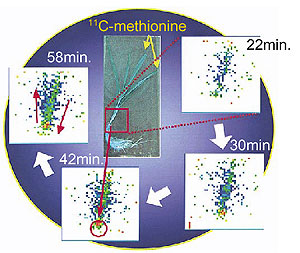
Methionine is the precursor of mugineic acid, which plays an important role in plants, such as barley, in the solubilization and uptake of weakly soluble Fe. However, although methionine is distributed through-out the plant, it was unknown where the source of the methionine required for the mugineic acid synthesis is located. Using PETIS, we have succeeded in imaging the translocation of methionine labeled with 11C, as shown in Fig. 9-11, and have found that the methionine used as the precursor of mugineic acid originates in the roots.
Using PETIS, information on plant responses to environmental change can be accumulated systematically from the observation of material translocation in plants placed under different environmental conditions. |