8-6 |
Nondestructive Evaluation of Spatial Distribution of Interlayer Structure in Multilayer X-Ray Mirrors Using X-Ray Standing-Wave |
 |
||
|
 |
||
|
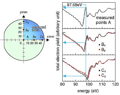
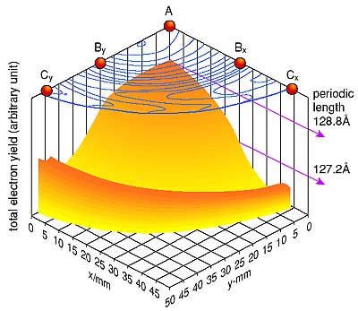
| Multilayer X-ray mirrors have been employed in high-flux X-ray optical systems for highly brilliant synchrotrons and laser-plasma X-rays. X-ray mirrors have two kinds of nano-meter-thickness thin films deposited alternately on a substrate several inches in diameter. In the development of such multilayer X-ray mirrors, evaluations of interlayer structure are necessary to improve their optical quality. The interlayer structure usually is evaluated by transmission electron microscopy (TEM). This is a destructive method because TEM evaluations require the multilayer X-ray mirrors be cut. To avoid this problem, we have demonstrated nondestructive evaluation of the spatial distribution of the interlayer structure in multilayer X-ray mirrors utilizing an X-ray standing-wave. In this method, the monochromatized synchrotron radiation beam is irradiated perpendicularly on multilayer samples. The resulting photocurrent generated in the samples is measured. When scanning the photon energy of the incident beam, the measured photocurrent drastically changes when the photon energy satisfies the specific optical condition (Bragg condition), which is dependent on the interlayer thickness (Fig. 8-13). Mapping measurements in the sample plane (x, y-axes) of the photocurrent (z-axis) around the Bragg condition can therefore nondestructively provide spatial information of the interlayer structure in the sample plane. The mapping spectrum of the quarter of the 4-inch-diameter Mo/Si multilayer X-ray mirror visually illustrates that the periodic-lengths of the Mo/Si layers gradually become shorter from the center toward the periphery (Fig. 8-14). This mapping measurement can be performed within one hour, which indicates that this is a less complicated evaluation of the interlayer distribution than conventional methods. This X-ray standing-wave method is expected to be utilized in the development of multilayer X-ray mirrors. |
| Reference Y. Muramatsu et al., Total-Electron-Yield X-Ray Standing-Wave Measurements of Multilayer X-Ray Mirrors for Interface Structure Evaluation, Jpn. J. Appl. Phys., 41, 4250 (2002) |
| Select a topic in left column |  |
| Persistent Quest Research Activities 2002 Copyright(c) Japan Atomic Energy Research Institute |