"Amyloid" diseases including various neuro-degerative diseases such as Alzheimer's disease and bovine spongiform encephalopathy (BSE, the so-called mad cow disease) share characteristics that misfolded proteins form filamentous structures called the amyloid fibrils and these fibrils are accumulated as deposits in tissues and organs. Recently, it has become known that not only the proteins related to the amyloid diseases but also a wide variety of proteins that are not related to any diseases form amyloid fibrils. Understanding the mechanism of the amyloid fibril formation is not only important for the development of therapeutic strategies against the amyloid diseases, but also is of considerable help to gain an insight into the generic properties of proteins.
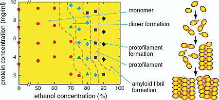
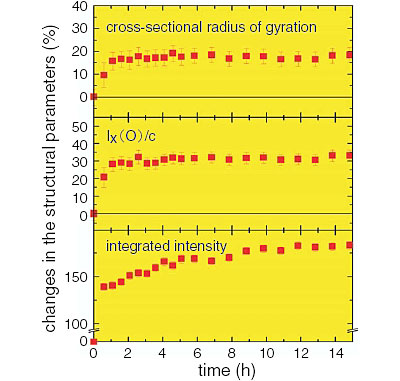
We employed hen egg white lysozyme (HEWL) as a model protein to study the mechanism of the amyloid fibrils and to investigate the process of the amyloid fibril formation of HEWL in ethanol solution with X-ray and neutron small-angle scattering. Small-angle scattering is an important method to study gross shapes and association states of particles in solution. We performed X-ray and neutron small-angle scattering experiments of HEWL in various HEWL concentrations and in various ethanol concentrations. From the results of these experiments, a phase diagram of the amyloid fibril formation was obtained, and it was shown that the amyloid fibril formation of HEWL occurs through the formation of the dimers, the formation of the protofilaments, and the formation of the amyloid fibrils by lateral association of the protofilaments (Fig. 4-6). We also investigated temporal changes of the structural states of HEWL in the process of the amyloid fibril formation with time-resolved neutron scattering. From the temporal changes of the structural parameters obtained, it was shown that association of the amyloid fibrils occurs following the formation of the amyloid fibrils by the association of the protofilaments (Fig. 4-7).
It is expected that understanding the mechanism of the amyloid fibril formation with neutron and X-ray small-angle scattering will ultimately contribute to understanding the mechanism of the amyloid diseases and their therapeutic strategy.
|