High-brilliance X-ray sources, representative of 3rd-generation synchrotron radiation, have been applied in various fields such as material sciences and biosciences, and are providing more novel research breakthroughs. Further, the development of highly coherent X-ray lasers as next-generation X-ray sources is now progressing. In our research group, we, as a world pioneer of this field, have succeeded in developing a highly coherent X-ray laser in the soft X-ray region. With it, the microscopic surface structures of the ferroelectric barium titanate (BaTiO3) were successfully observed with a picosecond (ps)-time resolution.
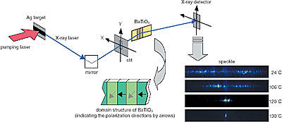
Traditionally, such microscopic structures are observed with Atomic Force Microscopy or Electron Microscopy. However, since the scanning time for these measurements usually takes several minutes or more, it is impossible to observe precisely microscopic structures having fast dynamics. Here then, we have succeeded in developing a new method to observe the microscopic structures with both a higher spatial resolution and a higher temporal resolution. The object sample is illuminated by the X-ray laser having a wavelength of 13.9 nm and a pulse width of 7 ps, which was developed at the Advanced Photon Research Center. The information of microscopic structure of the object then is recorded in the reflected interference pattern, i.e., speckle pattern, instantaneously within the time scale of the 7 ps of the X-ray laser pulse. The spatial resolution is about the order of the X-ray wavelength, 13.9 nm. Using this method, we have successfully observed the annihilation process of the microscopic surface structure of the ferroelectric domains of BaTiO3 as the temperature approaches the Curie temperature of this crystal (Fig. 5-4).
It is anticipated that this novel method can serve as a powerful tool for material science studies in the future.
|