An ion beam acts as a mutagen when it deposits a lot of energy locally and causes clustered damage in DNA. However, because there is no method to detect DNA damage with accuracy, it is not known how much DNA damage is generated by such an ion beam or where it occurs. Abasic site (AP site) is one of the most easily-induced and representative DNA damages, although there exist scores of other known defects, such as oxidation of base, DNA strand breaks, etc. ELISA and other methods have been developed to determine the number of AP sites, but there are no accurate methods to determine the location and extent of damage induced in DNA molecules.
In this study, we have succeeded in determining the location of single-molecule AP sites in DNA, using the combination of an aldehyde-reactive probe (ARP) that will combine with AP sites with nearly 100% efficiency and streptavidin conjugated with Cy5, a fluorochrome.
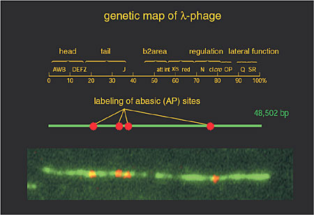
Fig. 6-12 shows a fluorescent-visualization of abasic sites that have been generated on lambdaDNA as a model DNA. First, AP sites were induced through treatment of the DNA with citrate. Then, the abasic sites were combined with biotinylated APR, after which the AP sites were fluorescent-labeled through reaction with Cy5-conjugated streptavidin. The number of fluorescent-labeled sites has been found to match the number of damaged sites detected by the ELISA method, indicating that AP sites have been labeled with an efficiency of approx. 100%. Four AP sites have been labeled with red color in Fig.6-12. The present technique is thought to be applicable to medical science, as in the investigation of mutations, cell death, and so on.
|