4-6

Investigating the Molecular Mechanism of Regulation of Muscle Contraction with Neutron Scattering
- Conformational Change of Troponin C within the Muscle Thin Filaments as Detected by Neutron Scattering -


Fig. 4-16 Structure of the muscle thin filament

In the thin filaments of skeletal and cardiac muscles, proteins called actin, tropomyosin, troponin I, troponin C, and troponin T, are arranged regularly to form a filamentous protein complex. The regulatory mechanism of muscle contraction lies within this complex.


Fig. 4-17 Method of sample preparation for neutron scattering measurements

To "see" only troponin C within the thin filaments, troponin C is first deuterated and incorporated into the thin filaments (a). By this treatment, the thin filaments containing troponin C, which have a density different from other components, are prepared. The density of the solvent in the sample solution is then matched to that of the components that are not deuterated by making a solvent containing the appropriate fraction of deuterated water (b). This treatment makes only deuterated troponin C "visible" to neutrons. From the neutron scattering measurements under this condition, scattering curves arising only from troponin C within the thin filaments can be obtained.


Fig. 4-18 Neutron scattering curves of troponin C within the thin filaments, and the results of the analysis

Clear differences were observed in the scattering curves of troponin C within the thin filaments with and without Ca2+. This indicates that troponin C assumes different comformation with and without Ca2+. Analysis by the model calculation showed that troponin C has an extended dumbbell-like shape, and when Ca2+ is present, it is elongated by about 0.2 nm and moves toward the axis of the thin filament by about 0.4 nm.


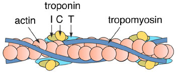
Muscle contraction occurs when the two main components of muscle, the filamentous protein complexes, "the thick filaments" and "the thin filaments," slide past each other. Elucidation of the molecular mechanism of muscle contraction and its regulation is one of the important challenges in biophysics. Muscle contraction is regulated by the Ca2+ concentration in the muscle cells. In skeletal and cardiac muscles, the regulatory mechanism lies in the thin filaments. In the thin filaments, five kinds of proteins, actin, tropomyosin, troponin C, troponin I, and troponin T, are arranged regularly (Fig. 4-16).
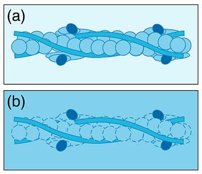
To understand the regulatory mechanism of muscle contraction by the thin filaments, it is very important to know the structure of each component within these filaments. As a first step, the structure of troponin C within the thin filaments was investigated by neutron scattering. Troponin C was deuterated, and then incorporated into the thin filaments. Neutron scattering experiments were performed on this sample under conditions where the components other than troponin C were rendered "invisible" to neutrons. The results of these experiments provided the structural information of troponin C within the thin filaments (Fig. 4-17). Such measurements can be done only with neutrons.
Analysis of the results of the neutron scattering experiments showed that troponin C within the thin filaments assumes an extended dumbbell-like shape, and by the binding of Ca2+, troponin C is elongated by about 0.2 nm and moves toward the axis of the filament by about 0.4 nm (Fig. 4-18). Combining these results with those of similar measurements of other components, which are now underway, should provide detailed information on protein-protein interactions related to the regulatory mechanism of muscle contraction.
This study was supported in part by the Special Coordination Funds from the Ministry of Education, Culture, Sports, Science and Technology, Japan.


Reference
F. Matsumoto et al., Conformational Changes of Troponin C within the Thin Filaments Detected by Neutron Scattering, J. Mol. Biol., 342 (4), 1209 (2004).

Select a topic in left column

Persistent Quest Research Activities 2004
Copyright (C) Japan Atomic Energy Research Institute