A dosimeter, which is used for monitoring of personal dose in routine radiation work, cannot accurately estimate dose in some cases involving extremely high-dose exposure, e.g., radiation accidents. In such unexpected cases, dose can be reconstructed based upon interactions of the human body with radiations. Electron spin resonance (ESR) measurement of tooth enamel in an exposed person is regarded as a useful technique because of the stability of the measured materials. This method focuses on the proportional relationship between the measured signal in ESR and dose to tooth enamel (enamel dose). On the other hand, doses to organs or tissues, which are sensitive to radiations, should be clarified in the estimation of radiation exposure. As the organs or tissues are mainly contained in a human torso and their elemental compositions differ from tooth enamel crystal, a significant difference can be seen between the enamel dose and the organ doses.
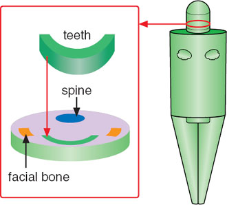
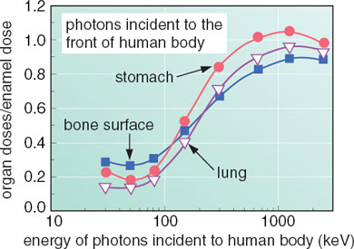
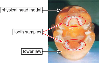
As a result, the relationships between tooth enamel dose and organ doses were clarified for external photon ( gamma- or X-ray) exposures. First, a human model was newly developed by incorporating teeth to analyze the tooth enamel dose by Monte Carlo calculations, as shown in Fig. 3-7. The numerical analysis using the new human model provided relationships between the tooth enamel dose and organ doses for exposures to photons with various energies and incident angles to a person. Fig. 3-8 presents a typical calculated result. In addition, the calculated results were verified by photon irradiation experiments with a physical human head model containing tooth samples, as depicted in Fig. 3-9.
The results obtained are essential to dose estimations by ESR measurements of tooth enamels and are adopted in some technical reports published by international institutes, e.g., IAEA. The data derived by this study, therefore, have been internationally utilized for retrospective dose assessments in some past events.
|