Studies on the molecular structure of actinide compounds in excited
electronic states are in progress through the use of resonance
Raman scattering. As shown in Fig. 7-1, resonance Raman scattering
(or resonance Raman effect) occurs when the exciting frequency
approaches or enters the region of electronic absorption of this
system. The Raman scattering intensity of the sample molecule
is then strongly enhanced as compared with that of ordinary Raman
scattering. Recently, such resonance Raman scattering has been
observed for uranyl nitrate (UO2(NO3)2), one of the well-known compounds in the actinide series. In
the electronic ground state, the uranyl ion ( UO22+) is linear and has three fundamental vibrations: asymmetric stretching,
symmetric stretching and degenerate bending. The uranyl ion also
has a strong electronic absorption band near 430 nm in the visible
region. It has been found that the resonance Raman effect occurs
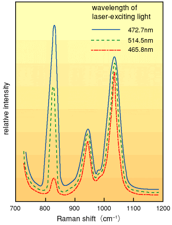
in the symmetric vibration ( O-U-O O-U-O ), observed at 835 cm-1. The Raman scattering intensity changes markedly depending upon
the wavelength of the laser-exciting light as shown in Fig. 7-2.
From the analysis of exciting-light dependency of the symmetrical
vibration, the conclusion can be drawn that the uranyl ion in
the excited electronic state has an equilibrium conformation linearly
distorted along the symmetric stretching mode and its bond distance
lengthens by approximately 0.014 nm in the direction of the uranyl
(O-U-O) axis. ), observed at 835 cm-1. The Raman scattering intensity changes markedly depending upon
the wavelength of the laser-exciting light as shown in Fig. 7-2.
From the analysis of exciting-light dependency of the symmetrical
vibration, the conclusion can be drawn that the uranyl ion in
the excited electronic state has an equilibrium conformation linearly
distorted along the symmetric stretching mode and its bond distance
lengthens by approximately 0.014 nm in the direction of the uranyl
(O-U-O) axis. |