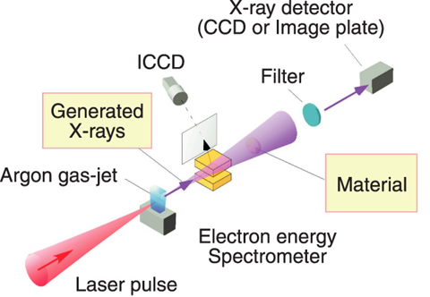
Fig.4-30 A schematic of laser-plasma X-ray generation device
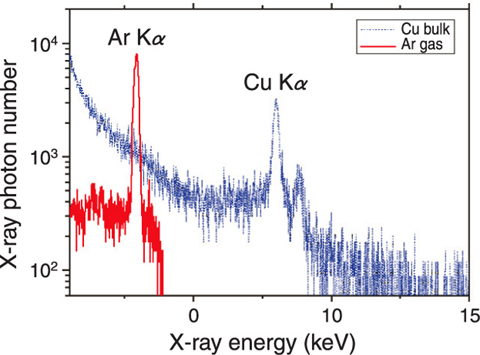
Fig.4-31 Obtained X-ray spectrum (red)
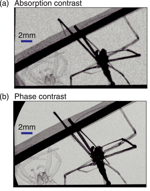
Fig.4-32 Absorption contrast (a) and phase contrast (b) imaging using the X-ray device
In X-ray imaging, the absorption contrast method is usually used, in which an image is formed by intensity variations due to absorption of X-rays in a material. However, it is difficult to observe soft tissue or small animals clearly and in detail because of their low absorption efficiency. Therefore, the phase contrast method, in which the change of refraction of X-rays in the material instead of absorption is detected, becomes an alternative. It requires X-rays having good spatial coherence. Thus, a specially designed X-ray tube, a synchrotron light source from a large-size accelerator, or laser-plasma X-ray source are used for the purpose.
However, the X-ray tube and laser-plasma X-ray source are small but less bright, while the synchrotron radiation source is bright but huge, therefore a new X-ray device which is bright and compact is demanded.
In this study, we have developed such a new device by optimizing the conditions of laser irradiation. In a laser-plasma X-ray source, a laser irradiated on materials produces plasma, and then the accelerated electrons excite ions, which radiate X-rays through atomic transitions. The previous X-ray source produced some undesirable Bremsstrahlung, and thus the desired X-ray had low brightness. We succeeded in maximizing the X-ray photon numbers in the required spectral region by reducing the prepulse which is large in high intensity lasers and by controlling the focus position. These measures are effective in reducing undesired Bremsstrahlung X-rays, because the appropriate energy is thereby transferred to electrons. As shown in Fig.4-30 and Fig.4-31 Terawatt, 70 fs pulse width Ti: sapphire laser pulses were focused onto an Ar gas jet. We obtained high-contrast X-rays; that is, undesirable X-rays were suppressed and the desired X-rays had a higher intensity, as seen in Fig.4-31. This new X-ray source enables us to reduce the total X-ray dose on a material or living body and obtain sharp images. In addition, the peak brightness is 1020 photons/s/mm2/mrad2, which is comparable to that of the synchrotron radiation. Because the pulse duration of this source is estimated to be on the order of 100 fs, it can be utilized to measure ultrafast phenomena.
We attempted absorption and phase contrast imaging of spiders with this source (Fig.4-32). Fine structure that cannot be seen in the absorption contrast image can be seen in the phase contrast image. Thus, we have demonstrated that this new X-ray source is suitable for phase contrast imaging.
This X-ray source produces spatially coherent, high brightness, high contrast X-rays. When this device is used in phase contrast computed tomography, it allows us to observe fine structure and reduces undesirable background X-ray dose to patients, and so this device is expected to be a useful new medical tool.

