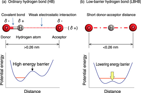
Fig.4-21 General aspects of an ordinary hydrogen bond (HB) and a low-barrier hydrogen bond (LBHB)
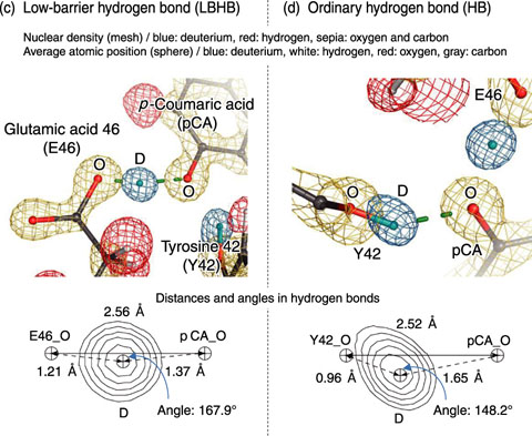
Fig.4-22 LBHB and ordinary HB in photoactive yellow protein (PYP)
Hydrogen bonds (HBs) play an important role in the stabilization of protein structures and in enzymatic catalysis. We first demonstrate that a particular kind of HB, low-barrier hydrogen bond (LBHB) (Fig.4-21), exists in a protein and regulates protein function.
The LBHB has been reported to be formed in organic molecules in several specific conditions, e.g., under high pressure or in crystalline state. The formation of LBHBs in proteins was proposed in the early 1990s. However, since there have been no direct observations of LBHBs in proteins, it has been a matter of debate. To identify an LBHB directly, it is essential to observe the position of the hydrogen (H) atom. We succeeded in demonstrating the existence of an LBHB in a photoreceptor protein, photoactive yellow protein (PYP), by using high-resolution neutron crystallography. This analysis identified 819 H positions and showed the existence of an LBHB (Fig.4-22). Since crystallization buffers were prepared with heavy water, H atoms involved in HBs were replaced by deuterium (D) atoms. A D atom is located near the point midway between the phenolic oxygen (O) atom of the photon-absorbing chromophore p-coumaric acid (pCA) and the carboxylic O atom of glutamic acid 46 (E46) of PYP. This deuterium atom is not covalently bound to either of the two O atoms. This experimental result provides direct evidence of the HB being an LBHB. It is known that the interatomic distance between the two O atoms increases after photon absorption by pCA. In such a situation, the LBHB cannot be formed. In this study, we propose a novel mechanism for photon signal transduction: upon the conversion of the LBHB into an ordinary HB, a light signal is transmitted from the chromophore to the protein. In addition, the present neutron crystallographic analysis indicates that the prerequisites for LBHB formation are that the donor-acceptor distance should be small and the donor and acceptor should have similar pKa values.
The present results show that neutron crystallography, which can directly identify H positions, is an important technique that can help improve our understanding of the molecular mechanism of protein function. The conversion between the LBHB and the ordinary HB has the potential to regulate protein function. The creative use of these HBs is expected to be a new guideline of drug design.

