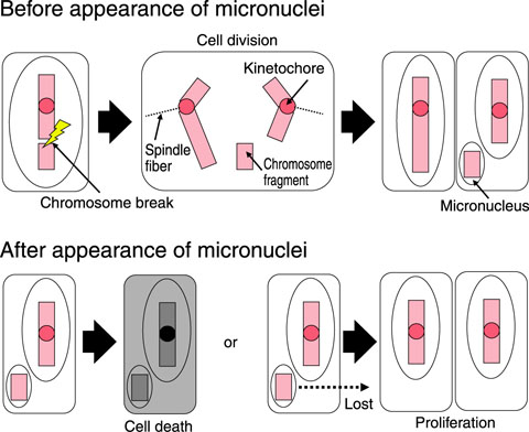
Fig.4-25 Appearance of micronuclei and their fate
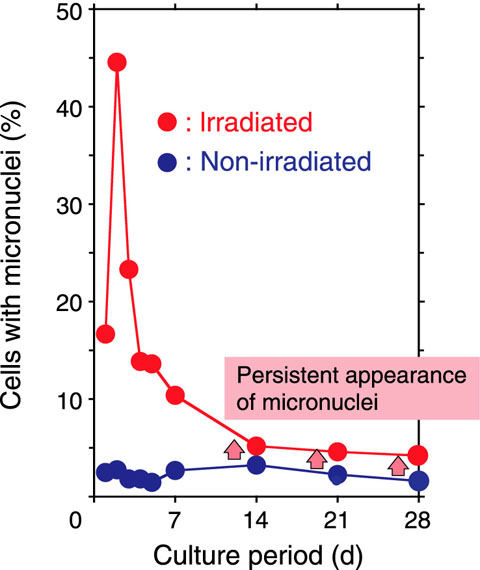
Fig.4-26 Persistent appearance of micronuclei after irradiation
Plants have chromosomes consisting of DNA and protein in their cell nuclei. When ionizing radiation (IR) and oxidative stress induce chromosomal aberration such as micronuclei (MN) (Fig.4-25), the genes corresponding to the chromosomes with aberrations undergo mutation. Recent studies have found evidence for the persistent occurrence of chromosomal aberrations in the descendants of irradiated mammalian cells. We thus cultured irradiated plant cells and measured the frequency of micronucleus induction in order to examine chromosomal aberrations in the descendants of irradiated plant cells.
In our experiments, tobacco cultured cells were irradiated with 40 Gy of 60Co γ-rays. Irradiated and non-irradiated cells were cultured in discrete vessels and aliquots of proliferated cells were transferred into new vessels every 7 days. Furthermore, a fraction of the cultured cells was collected to determine the number of cells and MN.
Irradiated cells as well as non-irradiated cells actively proliferated and the number of irradiated cells increased 223-fold in the investigation period. In irradiated cells, the fraction of cells with MN reached the maximum on day 2 and decreased with cell proliferation from day 3. It reached a minimum on day 14 and later, being greater than non-irradiated cells by almost twofold (Fig.4-26). From the fact that MN disappear soon, it is certain that MN persistently appeared after 20 or more cycles of cell division in the irradiated tobacco cells. It is supposed that oxidative stress is induced in the descendants of irradiated cells via an unknown mechanism and that MN appear persistently since IR enhances oxidative stress in the irradiated cells, which in turn induces chromosomal aberration.
In summary, we found that MN persistently appear in the descendants of irradiated plant cells. Cells with MN finally lose their genetic information, partly because MN are not inheritable. We thus point out that genetic information of irradiated plants can be altered later as well as immediately after irradiation. It is necessary to obtain a good understanding of the significance of the persistent appearance of chromosomal aberration for mutation breeding of plants using IR.

