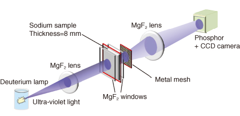
Fig.5-31 Set-up of the imaging experiment
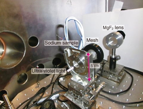
Fig.5-32 Photograph of an 8-mm-thick sodium sample and the imaging system
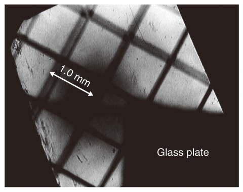
Fig.5-33 Image of a metal mesh composed of 100-μm-thick wires spaced by 1 mm, illuminated by ultra-violet light that shone through an 8-mm-thick sodium sample
The surface of fresh sodium reflects visible light. As is well known, surface electrons oscillate when exposed to incident light and emit light at the same wavelength. At shorter wavelengths of incident light, the electrons cannot follow the electric field of the light, and the light penetrates the sodium interior. However, the light is significantly dumped by phenomena such as electron-phonon scattering. To date, transparent sodium has not been experimentally investigated.
On the other hand, the optical properties of alkali metals (including sodium) have been intensively studied since the pioneering work of Wood in the1930s. In the1960s, the Oak Ridge National Laboratory team obtained a plasma wavelength of 218 nm. At wavelengths below 218 nm, sodium is partially transparent down to some threshold; namely, the core electron excitation wavelength (40.5 nm). The Oak Ridge team tried to measure the transmittance of a sub-μm-thick solid sodium layer coated onto a quartz and lithium fluoride (LiF) substrate. However, the thickness restricts the dynamic range of the transmittance. Therefore, we require a considerably thicker sodium region to clarify the transmittance.
In this study, we fabricate relatively thick sodium samples (>1 mm) in the spectral range beyond 115 nm, which corresponds to the shortest transmission wavelength of magnesium fluoride (MgF2) windows, and evaluate their transmittance. First, we determined the spectral transmittance between 115 nm and ∼200 nm. At ∼120 nm, the transmittance of 3-mm-thick solid sodium samples admitting the MgF2 windows was several tens of percent. Furthermore, the transmittance weakly depended on temperature up to 150 ℃ (note that the melting point of solid sodium is 97 ℃).
To confirm the transmittance, we also performed a simple imaging experiment with an 8-mm-thick sodium sample. Figs.5-31 and 5-32 show a schematic of the experiment and a photograph of the setup, respectively. We recorded a clear image of a metal mesh with 1 mm separation onto a two-dimensional charge coupled device detector (Fig.5-33). This result encourages us to design an optical imaging device for viewing objects inside or through solid or liquid sodium media, and to determine the physical origin of high transmittance, which has not previously been demonstrated.

