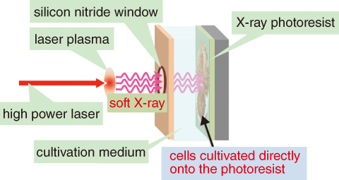
Fig.5-23 Conceptual design of the laser plasma soft X-ray microscope
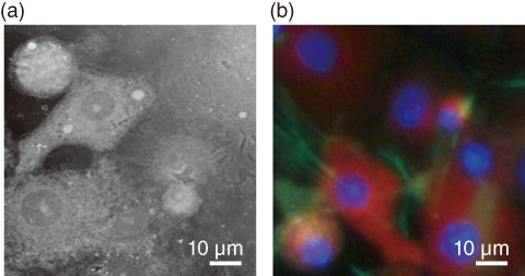
Fig.5-24 (a) Soft X-ray image and (b) fluorescent image of living cells
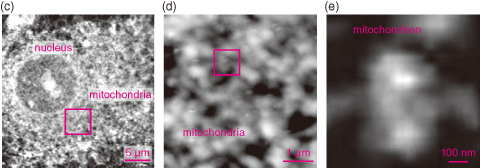
Fig.5-25 Soft X-ray images of the inner structures of living cells at different scales
We have developed a laser plasma soft X-ray microscope that combines a laser plasma soft X-ray source with contact X-ray microscopy. The laser plasma soft X-ray source offers high brightness and short pulse duration, whereas contact X-ray microscopy allows observation of cells cultivated in situ.
By combining these two techniques, we can observe living cells at a high spatial resolution. Intense soft X-rays were generated by irradiating a thin-foiled gold target with an intense laser beam. Fig.5-23 shows the conceptual design of the laser plasma soft X-ray microscope. Soft X-rays generated from the laser plasma were irradiated onto cells directly cultivated on an X-ray photoresist, and the soft X-ray images of the cells were recorded on the photoresist. The cells were then labeled with several fluorescent dyes and observed under a fluorescence microscope. The resulting fluorescent images were directly compared with the soft X-ray images.
Fig.5-24 shows the soft X-ray image (a) and the fluorescent image (b) of the living cells. The red, blue, and green regions in the fluorescent image are the mitochondria, nuclei, and cytoskeleton, respectively. Comparing the fluorescent images, we find that all organelles are clearly identified in the soft X-ray image.
Soft X-ray images of the inner structures of living cells are presented in Fig.5-25. Panel (c) clearly shows the mitochondria surrounding the nucleus. The mesh-like structures of the mitochondria are visible in panel (d), while panel (e) reveals a single mitochondrion, the first image of this organelle in living cells. The laser plasma soft X-ray microscope is expected to contribute further to life science studies.
The present study was partly supported by Japan Society for the Promotion of Science (JSPS) KAKENHI Grant-in-Aid for Scientific Research (C) (No.25390134).

