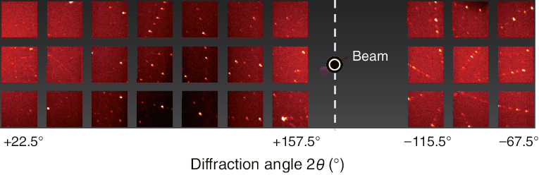
Fig.5-3 Neutron-diffraction pattern from an EuGa4 single crystal
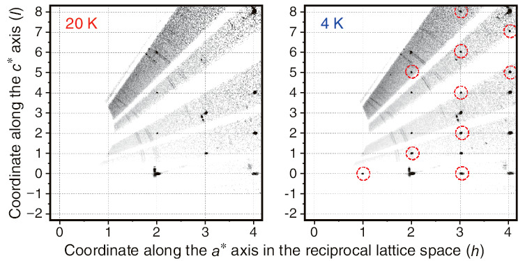
Fig.5-4 Neutron-diffraction intensities on particular planes at 20 K and 4 K
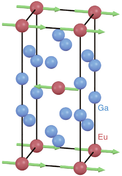
Fig.5-5 Crystal and magnetic structures of EuGa4 at 4 K based on analysis of pulsed-neutron-diffraction data
Magnetic-structure analysis by single-crystal neutron diffraction is an essential tool for studying the magnetic properties of materials. However, analysis of materials containing rare-earth elements such as Europium (Eu), Samarium (Sm), and Gadolinium (Gd) is not easy. As these elements show extremely high neutron absorption and non-monotonic neutron-energy dependence of absorption, the correct diffraction intensity cannot be obtained directly from the measured data. A correction method for such data is required.
In the present study, we developed an absorption-correction method for the single-crystal neutron-diffraction data from materials with high and complicated neutron absorption. By adopting this method for analyzing the high-quality neutron-diffraction data measured using the Extreme Environment Single Crystal Neutron Diffractometer SENJU, the magnetic structure of EuGa4 was successfully obtained.
Fig.5-3 shows the diffraction pattern from the EuGa4 single crystal measured by the detectors of SENJU. Although a shadow appeared due to the high neutron absorptivity of the sample, each diffraction spot was clearly observed. In general, the arrangements of the atoms and spins in materials can be obtained from the intensities of the spots. However, in the present case, the intensities extracted from the data must be corrected to suppress the effect of absorption before quantitative analysis of the structures. Therefore, an intensity correction based on the neutron-energy dependence of absorption for elements in the nuclear database was adopted for this analysis. The positions of the diffraction spots are as important as their intensities. Fig.5-4 shows the intensity distribution in the particular plane that reflects the tetragonal crystal lattice of EuGa4. The possible spin arrangements can be estimated from the positions of the spots appearing below 16 K, the ordering temperature of the spins in EuGa4. From these data, we conclude that the spins in EuGa4 are arranged alternately, as shown in Fig.5-5, and that the magnitude of the magnetic moment is ideal for divalent Eu.
We will develop the present analytical method of neutron-diffraction data more extensively, together with instruments for understanding the structure and properties of rare-earth compounds.
<Previous: 5 Applied Neutron and Synchrotron-Radiation Research and Development | Next: 5-2>

