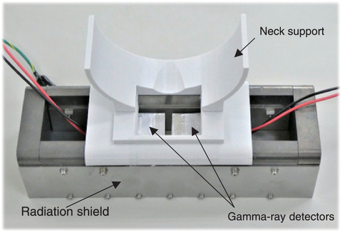
Fig.2-4 Developed thyroid dose monitor
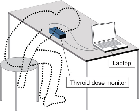
Fig.2-5 Subject's position during measurement
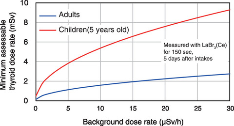
Fig.2-6 Minimum assessable thyroid equivalent dose
After a severe nuclear accident, the radioiodine monitoring in thyroids should be carried out for residents and emergency workers in the radiologically affected area. In-vivo measurement methods using thyroid dose monitors have typically been employed to determine the radioiodine activity in the thyroid. Individual monitoring should be conducted as early as possible after exposure to assess the equivalent dose to the thyroid; for example, the 131I remaining activity in a human's thyroid decreases according to its short half-life of 8.02 days. However, most thyroid monitors currently used in medical facilities are large and heavy and thus not easily transportable to evacuation centers, where many people can be measured immediately following an accident. After the accident at TEPCO's Fukushima Daiichi nuclear NPS, NaI(Tl) scintillation survey meters, which are widely employed to measure the ambient dose equivalent rate, were used to screen the 131I exposure in children's thyroids. Under high dose rate background conditions, however, this method cannot always provide reliable data.
We therefore developed a desktop-type thyroid dose monitor that can be easily installed in evacuation centers and works well under high background dose rates. The monitor comprises two gamma-ray spectrometers embedded into a box-shaped radiation shield made of lead and tungsten heavy alloy, as shown in Fig.2-4. The radiation shield effectively reduces the background radiation from the environment and the rest of the body; thus, the gamma-rays originating from the subject's thyroid can be selectively detected. Depending on the expected measurement situation, two kinds of gamma-ray spectrometers can be used: CdZnTe detectors for a high-energy resolution model or LaBr3(Ce) detectors for a high-efficiency model. As subjects are measured in the posture illustrated in Fig.2-5, the required table and chair could be easily prepared at an evacuation center.
A prototype of the proposed thyroid dose monitor was validated in the photon reference field established at the Facility of Radiation Standards at the JAEA. The resulting minimum assessable thyroid equivalent dose under varying background dose rates produced by a 137Cs source is shown in Fig.2-6. These results confirmed that for both children and adults, even under high background dose conditions, the monitor can assess thyroid equivalent doses of <10 mSv, which is sufficiently lower than the level at which health effects study is required. Further, calibration and background correction procedures were established to ensure accurate measurements.
After commercialization, the developed monitors will be deployed to nuclear facilities and off-site centers in prefectures where such facilities are located. Such monitors may also be useful in the field of nuclear medicine, such as in radioiodine therapy.
This work was supported by the Nuclear Regulation Authority (NRA) of Japan under Radiation Safety Research Promotion Fund (No.JPJ007057).
(Sho Nishino)
<Previous: 2 Research on Nuclear Safety and Emergency Preparedness | Next: 2-2>

