
 |
Topics
Analytical Method for Visualizing the Distribution of Trace Uranium in the Environment Using Superconducting Technology
-Advancing the Elucidating on the Migration of Trace Elements in the Environment-
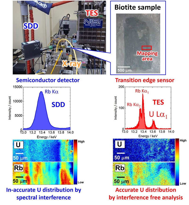
Fig. 1 Spot and mapping analysis of a biotite sample recorded using a conventional silicon drift detector and transition edge sensor
Uranium (U) is widely used as fuel for nuclear power generation worldwide. The migration behavior of U in the subsurface environment is important for assessing the safety of nuclear waste disposal. To understand U migration in the environment, it is necessary to analyze selectively the trace amounts of U distributed in environmental samples. Micro X-ray fluorescent spectroscopy is widely used for analysis of U in environmental samples. However, environmental samples often contain elements, such as rubidium (Rb), that interfere with U analysis. Hence, it is essential to develop a new analytical technique to detect the X-rays from only trace amounts of U in environmental samples.
In this study, we conducted X-ray fluorescence spectrometry analysis using a transition edge sensor (TES) which offer high energy resolution owing to utilization of superconducting technology. Through collaboration with universities and research institutes, the TES was successfully used as a detector in a synchrotron experiment with an X-ray microbeam. Results showed that the TES allows the accurate detection of the X-ray from trace amounts of U. Such detection could not be achieved with conventional solid-state detectors (SDD) due to interference of X-ray from large amount of Rb. Additionally, we successfully achieved the mapping analysis of the U in biotite, which revealed that U in biotite was distributed on the low Rb area in biotite.
Not only to U, TES can be applied to the analysis of other elements. Furthermore, TES enables the accurate determination of distribution of trace element at the ppm level even in environmental samples. We believe this new technique would greatly advance the migration study of various elements in the environment.
Acknowledgements
This work was supported by the Grants-in-Aid for Scientific Research (KAKENHI) from the MEXT (grant no. 18H05458, and 21H00162) and JSPS (grant no. 19H01145, 19H01960, 19K15606, 19K21893, 19K23432, 20K20527, 20K15238, 20K14524, 21H03585, 21H05443, 21K18649, 21K18917, 22F21313, 22H00166, and 22K18277). This study was performed with the approval of JASRi/SPring-8 (proposal no.2022A0174, 2022A0180, 2021A1610, 2019A1523, 2019B1498, and 2020A0174) and the Photon Factory (2022G126, 2020G670, 2020G081, and 2018S1-001).
Author (Researcher) Information
 | Name | Takumi Yomogida |
|---|---|
| Research Group for Nuclear Chemistry, Nuclear Science and Engineering Center |
Reference
Paper URL: https://doi.org/10.1038/s41467-024-45639-8
September 26, 2024
Nuclear Science and Engineering Research