
 |
Topics
Imaging Distribution of Trace Elements on Solid Surfaces
-A Highly Sensitive Imaging Method Using Laser Ablation and Isotopic Analysis-
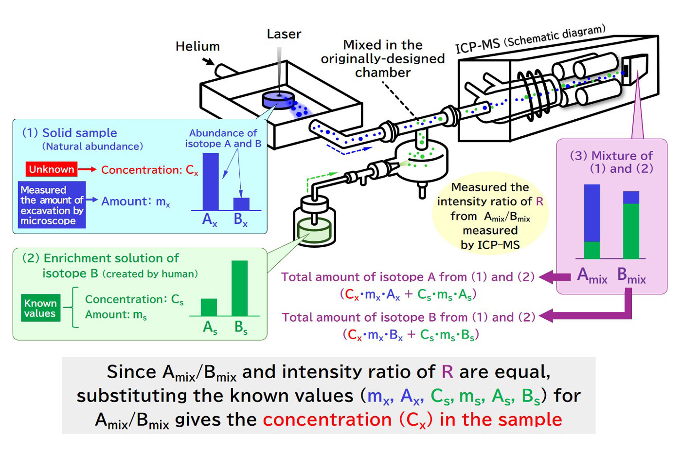
Fig. 1 Configuration of quantifying the imaging device and the theory behind the isotopic dilution method with mass spectrometry
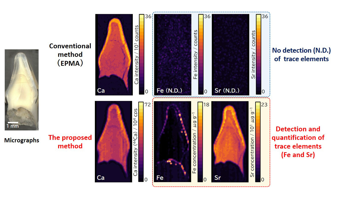
Fig. 2 Comparison of elemental imaging on a human tooth using the proposed method versus electron probe microanalysis (EPMA)
Proposed method determines the distribution of elemental concentrations from estimation of supplied amount of each isotope from the target sample through sequential mixing and measuring the aerosol containing isotopic standards with different isotopic ratios and known concentration, if the isotopic ratio and the amounts of the solid sample drilled by a LA could be provided. The isotopic ratio and the drilled amounts of the samples were determined by a LA (without mixing isotopic standard) and a digital microscope, respectively. As a result, both quantification and imaging of trace elements on the solid samples such as human teeth were achieved (Fig. 2).
The proposed method is promising for analyzing trace elements and RIs in a variety of biological and environmental samples.
Acknowledgements
This work was supported by JAEA Nuclear Energy Science & Technology and Human Resource Development Project Grant Number JPJA19H19210081, "Development of Methodology Combining Chemical Analysis Technology with Informatics Technology to Understand Perspectives Property of Debris and Tie-Up Style Human Resource Development."
Author (Researcher) Information
 | Name | Makoto Matsueda |
|---|---|
| Environmental Evaluation Research Group, Collaborative Laboratories for Advanced Decommissioning Science, Fukushima Research and Engineering Institute |
Reference
Paper URL: https://doi.org/10.1039/D3AN01028G
March 31, 2025
Research and Development Related to the Accident at TEPCO's Fukushima Daiichi NPS [R&D for environmental restoration]