In order to fabricate organic molecular devices as a complement to current semiconductor technology, new techniques are needed for doping polymer films with nano-size functional organic molecules. We have reported studies of a patterning method known as laser-induced molecular implantation technique or "LIMIT". This method successfully implants and positions fully functional organic molecules onto a polymer film using laser light activation.
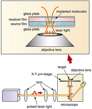
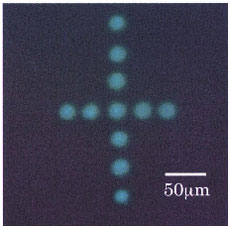
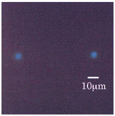
In the LIMIT method a polymer "source" film, which is doped with functional organic molecules, is irradiated with a pulsed laser while in close proximity (<1 micron) to a clean polymer "receiver" film. The dopant molecules are ejected from the irradiated part of the source film in a photo-thermal activation process and are transferred onto the target film where they become polymer dispersed. We have improved the spatial selectivity of this process by directing ablative laser excitation of the doped polymer film through the objective lens of an optical microscope (Fig. 4-8). In addition, we mounted an X-Y micrometer-stage on the microscope, so that by moving the stage we could vary the position of the target with respect to the pulsed laser light in order to create highly accurate spot patterning of functional polymer dispersed dopant molecules. In this way, precise positioning of functional dyes on a scale of a few micrometer is possible, allowing us to create larger periodic micrometer-scale structures. This process is demonstrated in Fig. 4-9, where a micrometer-scale cross shown. Figure 4-10 shows a fluorescence image of the minimum dot size obtained by optimizing the intensity of the pulsed laser light and the concentration of the dopant molecules in the source film. The minimum spot size was estimated to be about 4 micrometer.
This technique will be particularly useful for spatial arrangements of functional organic molecules for the fabrication of organic electric and optical devices, e. g. organic electroluminescent displays. |