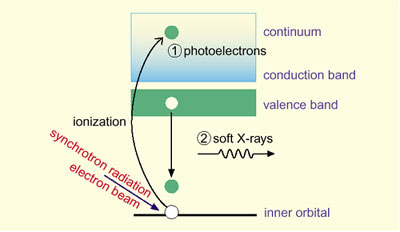
Since the early 1970's photoemission spectroscopy has been widely used for analyses of electronic structures and, in particular, it has played an important role in research on high Tc superconductors. Recently, a great deal of attention has been paid to soft X-ray emission spectroscopy using high-intensity synchrotron radiation. This kind of spectroscopy can reveal the individual components of atomic orbitals which make up the valence band in solid materials (Fig. 8-12). However, the emissivity is so small that it takes time from ~1 hour to 10 hours to obtain a good spectrum.
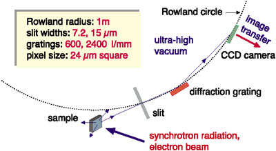
A spectrometer which we have developed employs a CCD camera as a detector (Fig. 8-13). The CCD surface consists of a few million pixels (24 micrometer square), each of which works as an independent counter for soft X-rays. To achieve sufficient energy-resolution (E/DeltaE:
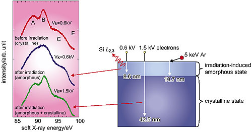
500 - 1500) and to cover a wide range of measurable energies (50 - 1500 eV), the soft X-rays are arranged to strike the CCD surface at a low angle (1.5 - 12°). Data obtained for pure silicon suggest that the distribution of atomic orbitals in the crystalline state is quite different from that in the amorphous state (Fig. 8-14).
The present work has brought about a substantial reduction in the measurement time, an extension of the measurable energies, and a reduction of the optical size. The spectrometer will be used for high-speed spectral measurements (i.e., a few seconds) by resonant excitation using synchrotron radiation and for detailed analyses of complicated electronic structures such as strongly-correlated electron systems. |