Water-soluble proteins are surrounded by hydration shells. In many cases, water molecules mediate biological processes via hydrogen bonds. However, in X-ray crystallography, the hydrogen atoms of water molecules are rarely observed even at ultra-high resolution. Recently, we have solved a high-resolution neutron crystal structure for myoglobin and for rubredoxin and its mutant. The intrinsic nature of the hydration shape for these cases has been found to be revealed in the course of this higher resolution analysis.
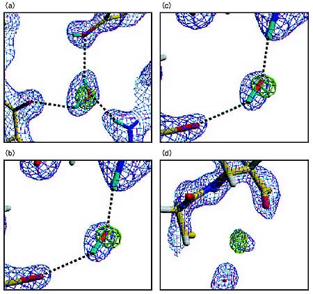
Water molecules adopt a variety of shapes in neutron Fourier maps (Fig. 4-13). We have categorized the shapes of water molecules into the following classes based on their appearance in Fourier maps: triangular, short ellipsoidal, long ellipsoidal and spherical. In the case of a triangular shape (Fig. 4-13(a)), the two D atoms and the O atom of the water molecule can be observed absolutely. For the short ellipsoidal peak (Fig. 4-13(b)), the oxygen position observed by X-rays is located at the end of the neutron Fourier peak, and only one D atom can be observed. We interpret short ellipsoidal peaks to represent water molecules rotationally disordered around their O-D bonds. For the long ellipsoidal peak (Fig. 4-13(c)), the O position is located in the middle of the neutron Fourier peak, and the two D atoms are only seen in part. The entire appearance is that of an elongated ellipsoid because of the molecular rotation or packing disorder around the axis parallel to the D-D axis. A typical example of the spherical shape peak is shown in Fig. 4-13(d). Only the center of gravity of this type of water molecule can be defined, because its orientation is totally disordered.
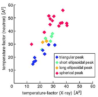
The dynamic behavior becomes more clear when the temperature-factors obtained by neutrons and X-rays are plotted as shown in Fig. 4-14. Here, it is remarkable to find that the small, intermediate and large temperature-factors correspond to water molecules having the triangular, short, and long ellipsoidal and spherical shapes in the neutron analysis, respectively. It can be concluded that the four major classifications of water molecules correspond closely with their dynamics.
The dynamics of water molecules revealed by the present analyses will be useful for computational analysis and for NMR analysis of the solution structures of proteins. |