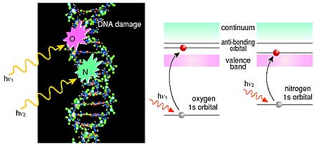
Studies employing synchrotron soft X-rays as probes to investigate genetic changes have highlighted biological effects such as mutations related with the molecular process of DNA damage. Site selective photoabsorption in a DNA molecule is a powerful technique to understand physicochemical mechanisms of DNA damage that induces radiobiological effects (Fig. 8-10). To study free radical species that cause complicated DNA damage, we have developed an EPR (electron paramagnetic resonance) system combined with a synchrotron soft X-ray beamline at SPring-8. The system enables us to investigate the radical process "in situ" in a DNA molecule during irradiation of soft X-rays.
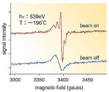
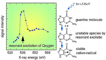
Fig. 8-11 shows typical EPR spectra of guanine (one of the DNA bases) at 77K. The spectra show formation of a short-lived unstable species clearly distinguished from a stable one that survives even after stopping the soft X-ray irradiation. The signal intensity of the short-lived species strongly depends on the soft X-ray energy; a characteristic large peak was found in the spectrum (Fig. 8-12). At the peak position (536 eV), the signal intensity was about five times stronger than that observed at 524 eV. It is inferred that the species is a radical cation formed as a result of the core level excitation of the 1 s electron in the oxygen of the guanine molecule to an anti-bonding molecular orbital (sigma*) of a carbon-oxygen bond following a resonant Auger decay in the molecule. Thus, we can infer a pathway for forming a final guanine lesion in DNA, as shown in Fig. 8-12. |