High-intensity pulsed-neutron sources using proton accelerators have been developed. A characteristic of these pulsed neutron sources is that peak neutron flux is a few hundred times stronger than that of constant-flux neutron sources from nuclear reactors, though the average neutron fluxes are the same and the neutron energies can be measured by the time of flight (TOF) method.
To perform neutron scattering experiments and neutron radiography under the optimum application of the pulsed-neutron sources, it is imperative to have neutron-imaging detectors with excellent performance for position resolution, time resolution, and counting rate, all of which were unavailable until now.
Therefore, we have examined pulsed-neutron imaging methods using phosphor/neutron converter detection sheets aiming at high-speed imaging using neutrons with high position resolution (Figs. 4-3 and 4-4).
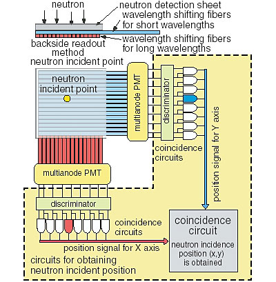
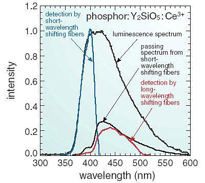
It is indispensable for the realization of the fast neutron imaging to shorten the luminescence lifetime of the phosphors in the neutron detection sheet. Therefore, Y2SiO5:Ce3+ with a lifetime of about 40 ns was used instead of ZnS:Ag phosphor, which has been used for neutron detection until now. It is necessary to develop new technology for the detection of the incident point of the neutrons because the luminescence rate of this phosphor is one twentieth that of ZnS:Ag, though the lifetime becomes shorter.
The backside readout method and the coincidence method were developed to improve the position resolution (Figs. 4-4 and 4-5). With the former method, the incident point of neutrons is obtained by detecting the photons emitted in the detection sheet with wavelength shifting fibers from the backside of the sheet. In the new method, the precise incident point is obtained by using the coincidence between these wavelength-shifting fibers.
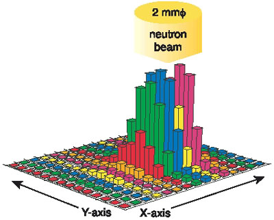
A neutron-imaging detector using a neutron detection sheet composed of Y2SiO5:Ce3+ as the phosphor and 7Li210B4O7 as the neutron converter was fabricated. The image detection experiments were conducted by using neutron beams in JRR-3. The image shown in Fig. 4-5 was obtained by measuring a 2-mm phi-collimated neutron beam. The position resolution is 0.6 mm. In addition, it was confirmed that a neutron image can be measured when neutrons of 3 Mcps arrive at the detector.
|