|
||
|
| Intense neutron fluxes are required to investigate the microscopic
structure in solid-state physics and biology. Neutron focusing is
difficult because a neutron has no electric charge and cannot be deflected
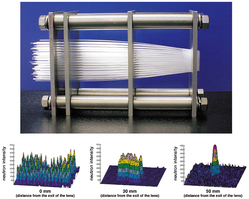
by magnetic field. To develop a focusing lens, we have applied the principle of external total reflection of neutrons incident on a smooth glass surface at a glancing angle which is caused by slow-neutron optical phenomena. Neutrons are transmitted through each inner-surface of the capillary, with multiple reflections. Many capillaries were assembled to point at a focal point, so that neutrons converge on a sub-millimeter region. This system is based on the principle of Kumakhov optics. Fig. 11-7 shows a series of images obtained near the focal point, which clearly confirm the focusing effect. The lens can be used for prompt gamma activation analysis in the study of elemental concentrations of the surface, and in neutron scattering for studying a material's structure and dynamics in an ultrahigh pressure environment using a sub-millimeter beam. |
| Reference
K. Soyama et al., Application of Multi-Capillary Fiber Neutron Microguide Tube, J. Nucl. Sci. Technol., 32 (1), 78 (1995). |
| Select a topic in left column |
| Persistent Quest-Research Activities 1997 Copyright(c)Japan Atomic Energy Research Institute |