 |
|||||
|
|||||
 |
|||||
|
|||||
@
 |
|||||
|
|||||
| Gadolinium-diethylenetriaminopentaacetic acid (Gd-DTPA) is used
as a contrast medium agent in magnetic resonance imaging (MRI)
for diagnosing blood vessel disease in the brain and the spine.
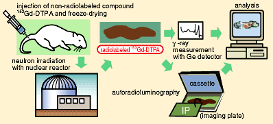
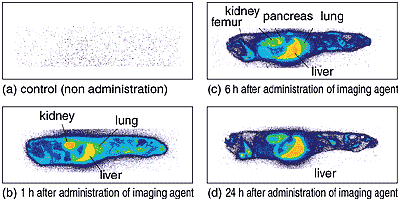
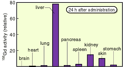
This agent is usually rapidly excreted after the diagnosis. We developed a novel, very highly sensitive method for detecting the residual distribution of imaging contrast agent in the whole-body. The agent, which is synthesized using gadolinium with the stable isotope 152Gd enriched to 30% from a natural abundance 0.2% (coexisting as 7 kinds of isotopes except 152Gd), was administrated to rats. After the rats were fed for predetermined periods, samples were prepared by means of freeze-drying them to be thin whole-body sections. When the animal samples were irradiated in a reactor, the 152Gd was turned to radioactive 153Gd by neutron reaction. The two dimensional imaging by autoradioluminography was carried out by measuring the radiation emitted from the samples (Fig. 4-10). Using the enriched isotope 152Gd, the detection sensitivity of this method was up to 150 times higher than the conventional method (Fig. 4-11). The residual distribution of 153Gd in the rat, namely the distribution of imaging contrast agent labeled with the 153Gd in each organ, was observed. It was found that the agent remained in the liver and kidney (Fig. 4-12). While the practice of the conventional method using radiolabeled 153Gd tracer has to be controlled by the radiation protection law from radiation hazards, the procedures of administrating the agent to animals and feeding them can be practiced in ordinary animal experimental facilities when the novel method with the enriched stable isotope is used. This method is highly appreciated in both domestic and foreign countries, especially in the United States, as promising wide application. |
| Reference K. Kobayashi et al., A High Sensitive Detection in the Whole-Body Residual Distribution of Imaging Control Agent Containing Gadolinium, Isotope News, 2, 12 (1998). |
| Select a topic in left column |
|
|
Persistent Quest-Research Activities 1998 Copyright(c)Japan Atomic Energy Research Institute |