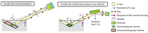
Fig.4-22 (Upper) Optics of strain scanning method
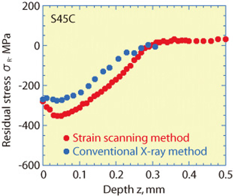
Fig.4-23 Distribution of residual stress measured by new type strain scanning method
To guarantee the reliability of structural component service, a non-destructive method is required to determine a precise distribution of the residual stress in materials. A strain scanning method based on X-ray diffraction method using synchrotron radiation is used to measure the strain and the stress. We improved this method, attempting to measure the residual stress of a local internal area of a material with high precision.
In order to measure the residual stress of a local area with the high precision, X-rays with high brilliance, high flux and high energy, and optics with high resolution are necessary. An original monochromator for high energy X-rays and an undulator in which permanent magnets are arranged in the straight section of the accelerator so that their south pole and the north pole alternate vertically were installed to obtain such X-rays. To achieve high resolution in the optics, a single crystal called an analyzer was installed (Fig.4-22). The analyzer is suited to measure with high resolution because it reflects monochromatized X-rays over a very small angle range, but the problem occurs that the X-ray intensity diffracted by the analyzer greatly decreases. Our proposed method enables a high space-resolution evaluation of the stress distribution which takes a few hours, whereas the conventional X-ray method takes several days (Fig.4-23).
The strain scanning method developed in this study can determine the stress distribution with the spacial resolution of several hundreds μm2 around a crack in internal material by using high energy synchrotron radiation X-rays. Furthermore in-situ stress measurement under high temperature or high pressure can be carried out because the measurement time of the strain scanning method is very short.
This newly developed method can be applied to various problems of concern to JAEA, such as stress corrosion cracking, development of materials for nuclear power generating plants, and the container for mercury target of "J-PARC". Furthermore, in the industry - academia - government collaboration, this method is applicable to research vital to the energy problem, for example, thermal barrier coating for a high efficiency jet turbine and solid oxide fuel cells.