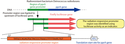
Fig.4-14 Identification of the radiation responsive promoter region of pprA gene
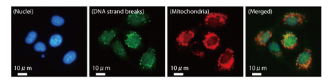
Fig.4-15 DNA strand breaks in mitochondria of mammalian cells following γ ray irradiation
Deinococcus radiodurans (D. radiodurans) possesses extraordinary resistance to ionizing radiation, which has been attributed to its highly proficient DNA repair capacity. We have identified the novel gene (pprA gene) from this bacterium, and revealed that the gene product (PprA protein) preferentially binds to DNA strand breaks and possesses DNA repair promoting activity. However, the radiation responsive DNA region (promoter) which is essential for the pprA gene expression has not been located. Moreover, though visualization of DNA damage utilizing the ability of PprA protein is promising, this detection technology has not been established.
To identify the radiation responsive promoter region, DNA fragments which may include the pprA gene promoter were ligated upstream of firefly luciferase gene. The luciferase activity was then monitored following γ irradiation as an index of gene promoter activity. It was revealed that the radiation responsive promoter region was located between positions -57 and -22 upstream from the pprA gene (Fig.4-14). Identification of the radiation responsive promoter is very useful finding to delineate the DNA repair network mechanism of D. radiodurans.
In mammalian cells that are sensitive to ionizing radiation, several DNA strand breaks result in cell death. Therefore, it is important to assess intracellular distribution and generative frequency of DNA strand breaks directly for the evaluation of radiation effect in mammalian cells. Certain DNA strand break visualization methods using proteins in mammalian cells associated with their own DNA repair have been developed. However, these methods have disadvantage that is difficult to detect initial DNA damage immediately following γ irradiation. To overcome this disadvantage, we developed a new detection method to visualize the initial DNA damage by utilizing the D. radiodurans PprA protein, which possesses an ability to bind to DNA strand breaks specifically. In this method, initial DNA damage can be visualized using fluorescence-labeled anti-PprA antibody bound to PprA protein (Fig.4-15). This detection method of DNA strand breaks with increased sensitivity will be useful in evaluating radiation effect in mammalian cells, and widely applicable to genotoxic tests in environmental and pharmaceutical fields.