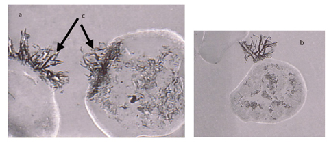
Photo 6-1 Transmission electron microscopy photographs of yeast cells exposed to a uranium solution (a, b)
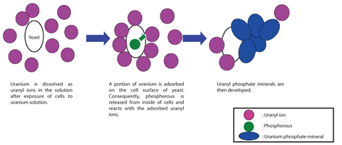
Fig.6-15 Schematic diagram of mineralization of uranium by yeast
"Yeast" is a single-celled fungus which multiplies by budding, associated with wine, beer, Japanese sake, and bread, but there is more to this organism. We have been conducting research to elucidate the interaction mechanisms of heavy elements with microorganisms. Through this research, we found that a strain of wine yeast mineralized uranium on the cell surface. We clarified the mechanism of the mineralization. Although this strain of wine yeast is presently just waste after the brewing of wine, it can used for long term confinement of uranium contained in naturally occurring radioactive material (NORM).
On the cell surface of yeast, adsorbed uranium reacts with phosphorous released from inside the cells. Consequently, uranyl phosphate minerals develop on the cell surface. It is known that uranyl ions are precipitated on the cell surface of microorganisms. However, the mechanism of the precipitation is not fully understand. We carried out an experiment exposing the cells of a strain of wine yeast to a uranium solution. We monitored change of uranium concentration with exposure time. The results showed that some uranium accumulated on the yeast cells. We analyzed yeast cells by transmission electron microscopy (TEM), and found that uranium bearing materials had developed on the cell surface (Photo 6-1). TEM, electron diffraction patterns and UV visible spectrometry indicated that uranyl phosphate minerals had developed on the cell surface, and that part of the minerals were in the intracellular region. On the contrary, no mineral was distinguished on the cells grown in a phosphorous starved medium. These results strongly suggest that uranyl phosphate minerals are formed at the cell surface by the reaction of the adsorbed uranium with phosphorous released from inside of the cells (Fig.6-15). Interestingly, concentrations of uranium and phosphorous and pH were below saturation levels for formation of uranyl phosphate minerals. These results indicate that cell surface creates specific conditions different from the bulk solution, where local saturation generating the minerals is attained.
Yeast is a eucaryote whose DNA sequence analysis has been completed. We are planning to identify the proteins which accumulate uranium in order to elucidate the accumulation mechanism of uranium at the molecular level.
<Previous: 6-7 | Next: 7 Nuclear Science and Engineering Research >