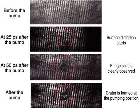
Fig.4-8 Soft X-ray interferogram of platinum surface pumped by ultra-short laser pulse
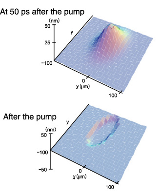
Fig.4-9 Retrieved surface profile from the interferogram
The dynamics of photo-induced phenomena, namely laser-induced melting, annealing, ablation, and ripple formation on solid surfaces, is the central issue in photon-aided advanced technologies for nanometer-scale fabrication. Photo-induced phase transitions, as well as photochemical and biological processes in nanometer space, also have to be clarified from a dynamic point of view. These phenomena often accompany changes in surface morphology proceeding in a very short (nano- to femtosecond) time scale, and thus are hard to explore using ordinary experimental techniques. The nonreversible and nonrepetitive nature of these phenomena adds more difficulties to time-resolved observation. In spatial resolution, it is directly related to the wavelength of the probe, and drastic improvement in resolution is being sought in extreme ultraviolet and soft X-ray regions by utilizing new light sources to investigate the structures of biological cells, magnetic domains, nano-particles/tubes, and fabricated devices.
The laser-driven plasma soft X-ray laser is an attractive light source for this purpose, because it has outstanding properties such as high monochromaticity, coherence, and short duration. We developed a pump-and-probe X-ray laser interferometer under the collaboration with Institute of Solid State Physics (ISSP) and University of Tokushima. The spatially coherent 13.9 nm laser illuminates a sample at a grazing angle of 24 degrees. The sample image is then transferred onto an X-ray CCD by a concave imaging mirror with a magnification factor of 21.3. The soft X-ray from the sample is divided into two parts by a double Lloyd’s mirror placed between the imaging mirror and the CCD, and is focused on the CCD with a small angle to produce interference fringes. The spatial resolution of this interferometer reaches 1 μm for lateral directions and 1 nm in depth, and the temporal resolution is the duration of the X-ray laser (~7 ps). This performance is sufficient for observing the surface dynamics of the initial stage of laser processing.
Using this interferometer, we demonstrated observation of the surface dynamics of a platinum (Pt) sample pumped by a 100 fs-duration infrared laser pulse. Fig.4-8 and Fig.4-9 show the temporal evolution of the interferogram of the Pt surface and the retrieved surface profile, respectively. After irradiation, a very small but substantial deformation starts at around 25 ps. At around 50 ps, apparent expansion of the surface is shown and the height of the peak reaches 30 nm, and finally a crater is generated. This is the first observation of the initial stage of laser processing or laser welding, and the result provides useful information to deep understanding of the fundamental processes in the laser-matter interaction. Our next objective is to improve the lateral resolution to ~100 nm by replacing the imaging mirror with a Fresnel zone plate.

