In order to develop a Very High Temperature Reactor (VHTR), which is a promising candidate for a Generation IV nuclear energy system, studies are being carried out on the technology for lifetime extension of in-core graphite and for the application of ceramic components. It is important to evaluate irradiation-induced change in the properties of graphite materials, such as dimensional change, Young’s modulus, etc., for lifetime assessment of the graphite components. These properties are strongly dependent on irradiation-induced change in the graphite microstructure (crystal grains and pores). Carbon fiber reinforced carbon composite (C/C composite) is made of carbon fibers impregnated with resin through a baking process in fabrication. It is one of the candidate materials for ceramic components of the VHTR. C/C composite contains many micro voids, cracks, and pores generated in the fabrication process, which strongly influence its material properties. It is thus necessary to develop an evaluation method for the properties of the material based on its microstructure.
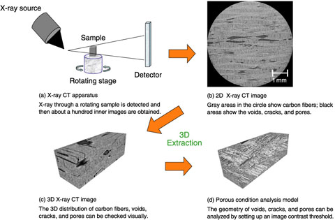
A new study has just been started to try to correlate the microstructures of graphite and C/C composite to their material properties by means of image-based modeling using X-ray computed tomography (X-ray CT), in cooperation with Toyo Tanso Co., Ltd. In this correlation method, the size and distribution of crystal grains and pores are quantified in micro regions by three-dimensional (3D) X-ray CT images and the material properties are then evaluated. As the first step of this study, the applicability of image based modeling with 3D X-ray CT images (Fig.8-12) was investigated for the C/C composite, provided by Toyo Tanso. To evaluate the porous condition from the X-ray CT images, it is necessary to binarize the images to distinguish between solid elements and pores. An analytical model was successfully developed through detailed X-ray CT image analysis and by setting appropriate threshold values for image binarization. Moreover, it was confirmed that this method can be also applied to graphite material.
The model correlating the microstructures of graphite and carbon material to their properties is now being developed using the above analytical method. This methodology is expected to enable us to predict irradiation-induced change in the properties of graphite materials, and to evaluate the lifetime of graphite components. It is also useful for the design of new graphite and carbon materials, and could greatly contribute to the general industrial field.