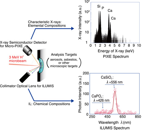
Fig.13-24 Schematic illustration of ILUMIS system combined with the micro-PIXE system (left) and spectra of PIXE (right upper) and of IL (right lower)
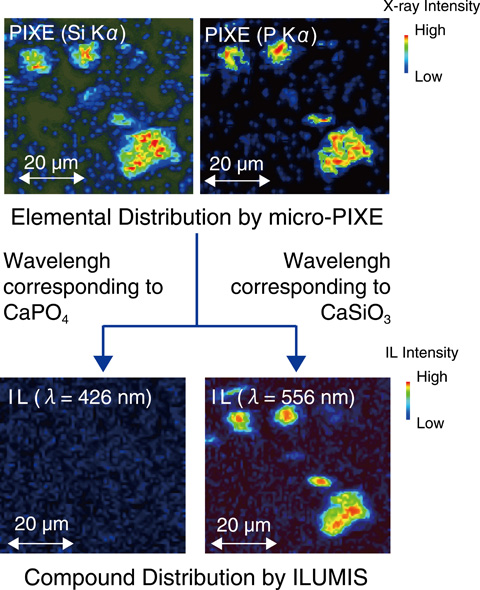
Fig.13-25 Examples of microscopic imaging of the elemental distribution obtained by PIXE (upper) and the chemical-state distribution by IL (lower) from aerosol samples with the scanning ion microbeam
Air-borne particles (aerosols) are abundantly floating in the atmosphere, providing surfaces that can transport harmful chemical substances and microbes. Large-scale transmission and expansion of chemical and biological toxins is a global environmental issue. To study the adhesive nature of particulate surfaces for such toxic materials, we must be able to visualize distributions of not only elements but also their chemical states in micrometer-sized structures.
At the ion accelerator facility of TIARA in JAEA/Takasaki, Particle-Induced X-ray Emission analysis using an ion microbeam (micro-PIXE) has been developed and utilized to analyze elemental distributions in individual microscopic samples. Micro-PIXE analyses detect characteristic X-rays emitted from elements heavier than Mg during the scan of an ion microbeam on a sample with typical spatial resolution of 1 μm.
During PIXE attributed to the ionization of inner-shell electrons, low-energy photons are also emitted due to the excitation of outer-shell electrons in the target. Such photon emission, which is called ion luminescence (IL), is strongly related to the chemical state of the elements or molecules; thus, measurements of IL enable us to determine chemical-state on the same sample area in which elemental compositions are analyzed by micro-PIXE analysis at the same time.
As shown in Fig.13-24, we have newly developed a wavelength-dispersive IL analysis system named Ion Luminescence Microscopic Imaging and Spectroscopy (ILUMIS) to obtain the chemical state distribution from microscopic target. The system consists of a large optical lens for high detection efficiency and an electrically cooled photon sensor for high signal-to-noise ratios with a grating effective between 200 and 850 nm.
Results obtained using the combined system of ILUMIS and micro-PIXE is shown in Fig.13-25 for individual aerosols. The particulate samples were scanned over with a proton microbeam in the atmosphere. The elemental compositions of the particles were obtained for silicon and phosphorus by the micro-PIXE, as shown in the upper panel of the figure. On the other hand, chemical state of the target was clearly obtained by ILUMIS. The result shows that the particulate samples consist not of calcium phosphate (CaPO4) but of calcium silicate (CaSiO3), as shown in the lower panel of the figure. These results demonstrate that ILUMIS combined with micro-PIXE is a compact and powerful tool for analyzing and imaging chemical compositions of microscopic samples.
This research was partially supported by a MEXT/JSPS Grant-in-Aid for Young Scientists (B) No.24710097 and by a JAEA Grant-in-Aid for Exploratory Research.

