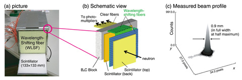
Fig.13-11 A high spatial resolution neutron image detector for iBIX
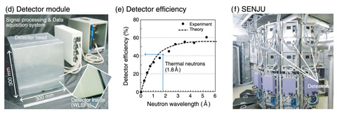
Fig.13-12 A large area neutron image detector for SENJU
Neutron diffraction on a single crystal of protein is a unique technique for identifying the hydrated position of the sample. However, acquiring high-quality data from the protein has inherent difficulties because the diffraction intensity is weak and scattered Bragg peaks are very close. A two-dimensional neutron detector that has a spatial resolution less than 1 mm and a detector efficiency higher than 50% with a minimum dead area is required. To fulfill such specifications, we have developed a detector that consists of scintillator and wavelength shifting fibers (WLSF) (Fig.13-11). With this detector, a neutron is detected through scintillation light. A spatial resolution of 0.9 mm is achieved by confining the scintillation light to a 0.4-mm thick scintillator and by reading the light using a 0.5-mm diameter fiber.
We also developed a ZnS/10B2O3 ceramic scintillator that has a four-fold larger neutron absorption cross section than a conventional ZnS/6LiF scintillator. The detector, which employs two scintillator screens and the dedicated photon counting electronics, exhibited a detector efficiency of 50% for thermal neutrons. This is higher than the conventional detector efficiency of 35%. The IBARAKI Biological Crystal Diffractometer (iBIX), populated with 30 such detectors, had a significantly reduced measurement time: only three days for a 1-mm3 protein sample. In contrast, more than a month was required with a conventional instrument built in the Japan Research Reactor-3 (JRR-3).
We have also developed a new image detector for the Single-crystal Neutron Diffractometer under Extreme Condition (SENJU) project. The aim of SENJU is to study crystal structures of materials under extreme environmental conditions, such as low temperatures and high magnetic fields. The SENJU project requires unique and unprecedented detectors that have a spatial resolution of 4 mm, high detector efficiency, and a large detector to scan a momentum space as efficiently as possible. We designed a dedicated detector head that incorporates a 1-mm diameter WLSF placed with a regular pitch of 4 mm (Fig.13-12). With this head, the detector exhibited a detector efficiency of 40% for thermal neutrons and the neutron-sensitive area was four times that of the iBIX detector. The SENJU shortened the measurement time to only a day for a small sample (1 mm3); this compares to a few days required for the measurement with a similar instrument in the JRR-3.
These new scintillation detectors significantly add to the high-performance neutron scattering instruments in the J-PARC. These new types of detectors would also be very useful in any application that requires measurement of the incident positions of neutrons.

