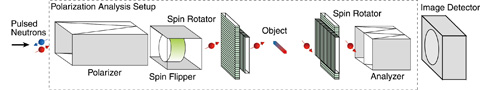
Fig.13-13 Schematic of magnetic field imaging system using polarized neutrons
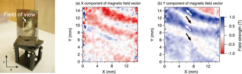
Fig.13-14 Observation of magnetic field distribution inside a soft magnetic foil
Because a neutron has a magnetic moment, it interacts with a magnetic field directly and precesses around the magnetic field. The resulting rotational angle of the neutron spin reflects the magnetic field strength and the direction in which the neutron moves. Therefore, by analyzing the neutron spin rotation at each position, we can obtain images of spatial distribution of magnetic fields. Because interactions between neutrons and magnetic fields depend on their velocities, measurements of the spin rotation as a function of velocity enable us to quantify the magnetic field. Thus, using pulsed neutrons is very suitable because they make available a wide range of neutron velocities; precision measurements can then be obtained by applying the time-of-flight method.
In this work, we developed a new technique for visualizing magnetic field distribution by combining the neutron polarization analysis with the pulsed neutron imaging method (Fig.13-13). Neutron spin rotation is recorded as the change in polarization at each position over a wide range of neutron velocities. In addition, by using spin rotators to select both incident and transmitted neutron spin directions, vector components of the magnetic field can be obtained; then the direction of the field can be determined. The images in Fig.13-14 show the magnetic field distribution of a soft magnetic foil obtained at the instrument “NeutrOn Beamline for Observation & Research Use (NOBORU)” of J-PARC MLF. These images contain stripe-shaped magnetic domains with magnetic field directions in neighboring domains being opposite one another (see arrows in Fig.13-14(b)). The magnetic field strength was evaluated to be 1.2 T. A high-flux neutron beam is required for analyzing the neutron polarization with fine spatial resolution. J-PARC’s high-intensity pulsed neutrons enabled us to not only visualize but also quantify the magnetic field with a spatial resolution of less than 1 mm.
The present study was partially supported by a JSPS Grant-in-Aid for Scientific Research (C), Grant No.22604009.

