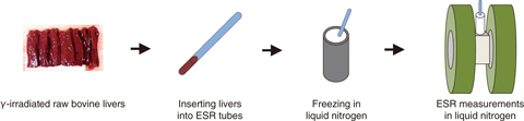
Fig.5-35 Schematic of electron spin resonance (ESR) measurements
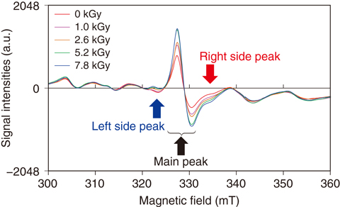
Fig.5-36 Changes of ESR spectra of irradiated raw bovine livers
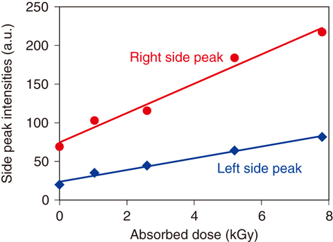
Fig.5-37 Dose responses of side peaks on bovine livers
To prevent foodborne illness in Japan, after the outside of raw meat had been roasted and then the cooked parts were trimmed off, the steak tartare must be prepared from only the inside meat in restaurants. However, since pathogenic bacteria such as some E. coli were found inside raw bovine livers where no disinfection method is effective, the sale of any raw bovine liver to eat as “liver-sashimi” has been prohibited since July 1, 2012. Providing for the radiation exposure that is used to sterilize bacteria out of a sealed food package without heating, detection methods have been developed to distinguish raw livers that have been disinfected by radiation.
At first, the irradiated raw bovine livers were subjected to the ELISA method, which was developed as for irradiated ground meats. The ELISA method could distinguish irradiated livers when such irradiation was done in the chilled condition (0 °C), but not at all could be detected when irradiation was done in the frozen condition (−80 °C).
The ELISA method can detect an oxidative lesion in DNA that reacts with active species (radicals) caused by ionizing radiation. Therefore, the radical migration to DNA might be inhibited in the frozen condition. Next, we tried to detect the radical itself. Basically, unpaired electrons of radicals can be measured using electron spin resonance (ESR) spectroscopy. This apparatus utilizes an electromagnetic wave similar to a microwave oven. Therefore, the method has a weak point in that the electron cannot resonate when the water within the samples absorbs the microwave. However, we have demonstrated that ESR can measure radicals in the flesh of fresh fruits irradiated as a disinfestation treatment for the plant quarantine. Thus, we thought that it might be possible to measure radicals in livers using ESR.
After raw bovine livers were irradiated in the chilled condition, they were measured at liquid-nitrogen temperature to suppress microwave absorption by stopping the motion of the water molecules during ESR measurement (Fig.5-35). According to the results, we found a main peak and two side peaks in the livers (Fig.5-36), and the side-peak intensities increased according to the increasing doses (Fig.5-37). Since the measuring errors were involved in experimental values, we assumed that the side-peak intensities of the raw bovine livers irradiated at approximately 3 kGy could be clearly distinguished from those that were not irradiated. Therefore, it is suggested that raw bovine liver treated by irradiation beyond 3 kGy can be distinguished using the ESR method.
Since the ESR method can be used to investigate the raw bovine livers being irradiated for disinfection, we believe that this technique can help consumers to choose reliable liver-sashimi.

