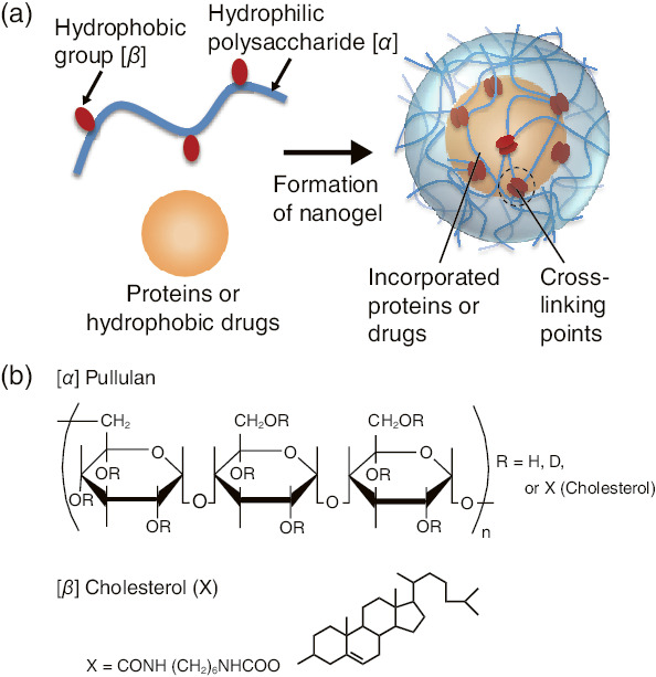
Fig.5-13 Formation of nanogels
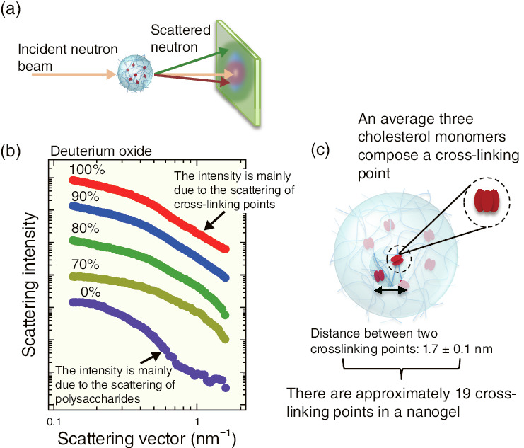
Fig.5-14 Small-angle neutron scattering of nanogels
Cholesterol-bearing pullulans spontaneously form stable nanogels of about 30 nm in diameter in aqueous solution due to hydrophobic interactions between cholesterol molecules (Fig.5-13). Because such nanogels possess a high loading capacity for proteins and drugs mainly through the hydrophobic interaction, they have been applied in drug-delivery systems. Although the spatial distribution of cross-linking points in the nanogels is closely related to their properties, no clear nanoscopic information has been obtained so far due to the complex structure with multiple components. This study aims to investigate the structure of the nanogels by means of contrast-variation small-angle neutron scattering (SANS), which enables us to determine the quantitative complex structure at the nanometer scale.
Small-angle scattering is a powerful technique that uses elastic-neutron scattering at a small scattering angle to study nano- to macro-structures (Fig.5-14(a)). The neutron-scattering length varies considerably between two isotopes; thus, the contrast-variation technique can be easily applied with neutrons by utilizing the difference in nuclear scattering arising from hydrogen/deuterium replacement in the system.
Fig.5-14(b) shows the scattering intensities of the nanogels dispersed in water with D2O fractions of 100%, 90%, 80%, 70%, and 0%. The scattering intensities vary with the D2O fraction in the water; this is characteristic of multicomponent systems because the contrast-matching points for pullulan and cholesterol against water are different. The partial-scattering functions for each component were individually extracted, after which the pullulan-network structure and the cholesterol distribution in the nanogels were evaluated. From our analyses, it was observed that the cross-linking points are formed by aggregation of trimer-cholesterol molecules, and that there are 19 cross-linking points in a nanogel.
The SANS experiments were performed using a time-of-flight diffractometer, TAIKAN at J-PARC, Japan. This study was supported partially by the Japan Society for the Promotion of Science (JSPS) KAKENHI Grant-in-Aid for Young Scientists (B) (No.25790087).

