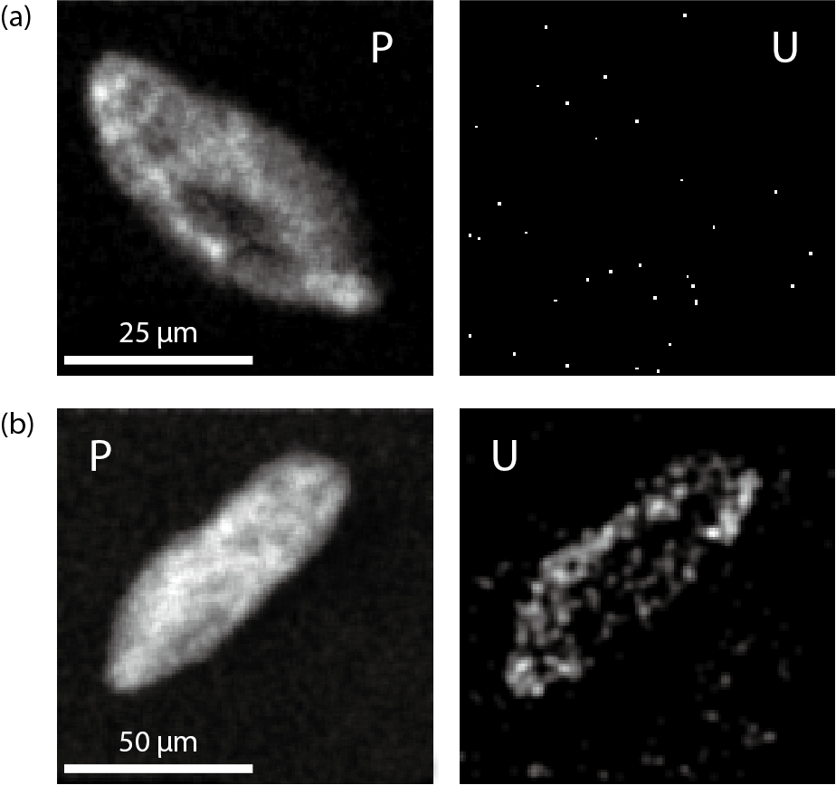
Fig.3-4 Adsorption of uranium on Paramecium cells
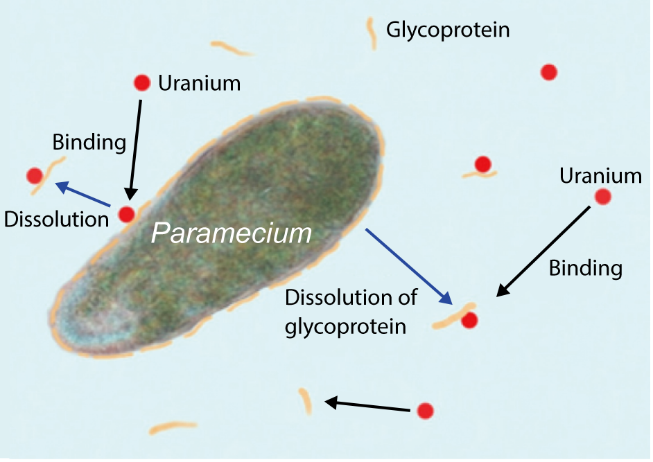
Fig.3-5 Binding of uranium to soluble surface glycoprotein of Paramecium cells
The migration of metal elements (including radionuclides) through geological and aquatic environments is presumably influenced by the biological functions of bacteria, such as adsorption on cell surfaces, phosphate mineralization, reduction, and oxidation. Environmental bacteria lie at the bottom of the ecological pyramid (food chain) in environments co-inhabited by themselves and larger creatures. Bacteria are consumed by protozoa (e.g. paramecia, ameba, and euglena), single-celled microorganisms with lengths of tens to several hundreds of micrometers. These tiny organisms control the bacterial populations. However, the roles of protozoa on the environmental migration of metal elements have not been previously investigated. To elucidate these roles, this study investigates the interaction between a representative freshwater protozoan (Paramecium sp.) and a metallic element (uranium).
Paramecium cells were exposed to aqueous uranium solutions in the laboratory. Uranium adsorbed on living cells was hardly detected using an element analytical device but was clearly detected on dead cells analyzed via micro-particle induced X-ray emission (micro-PIXE). Here, the Paramecium cells were killed using chemicals (fixatives) before running the experiment (Fig.3-4). After the experiment, we probed the cause of this large difference by analyzing the liquid phase. We found that a fraction of the uranium was bound to an organic substance released from living Paramecium cells. This organic substance was a large soluble glycoprotein with an approximate molecular size of 250 kDa.
The characteristics of the organic substance accorded with those of the glycoprotein covering the entire cell surface of Paramecium. This finding strongly suggests that dissolution of the uranium-biding glycoprotein from the living cell surface reduced the level of the adsorbed uranium to below the detection limit of the micro-PIXE instrument (Fig.3-5). Meanwhile, the uranium on the prekilled cells remained because the surface glycoprotein was chemically fixed to the cells.
Paramecium is sensitive to and poorly resistant to metal elements. Although the role of glycoprotein on the Paramecium cell surface is only partially understood, it may protect the cell surface from metal toxicity.
This work was supported by the Japan Society for the Promotion of Science (JSPS) KAKENHI Grant-in-Aid for Scientific Research (C) (No.25420910).

