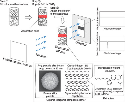
Fig.7-2 Experimental resonance neutron imaging method
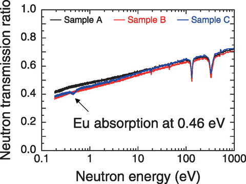
Fig.7-3 Measured neutron transmission
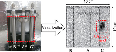
Fig.7-4 Photograph and neutron radiograph of the column
When reprocessing spent nuclear fuel, uranium (U) and plutonium (Pu) can be reused; all other elements are geologically disposed after vitrification. However, separating long-lived MAs from radioactive liquid waste is expected to reduce the volume and radiotoxicity of radioactive waste. Therefore, we developed extraction chromatography using a separation column filled with MA-compatible particles (i.e., CMPO-impregnated adsorbent). After the waste liquid is supplied, MAs form an adsorption band and can be separated from other elements due to their affinity to a CMPO extractant. In this manner, 99% of the MAs were separated from real waste liquid in the demonstration experiment. Our facing issue is the purity of recovered MAs, which must be improved for the nuclear fuel fabrication. We supplied europium (Eu3+) to the column as a simulated MA to observe the adsorption band by resonance neutron imaging for better understanding of the phenomena inside the column.
A schematic of the neutron irradiation experiment performed at J-PARC is shown in Fig.7-2. The column was irradiated with neutrons, and the transmitted neutrons were measured by a detector. The neutron energy was determined by the flight time of neutrons from the pulsed neutron source to the detector, and the nuclides and their spatial distribution were identified from the neutron absorption energy and the decrease in the number of neutrons after transmission, respectively. These data were converted to a neutron radiograph, which allowed us to observe the internal structure of the column without destroying it.
When light water (H2O) is used in neutron radiography, neutrons react with hydrogen, causing low transmittance and a low-resolution neutron radiograph. This problem was solved by using deuterium. The obtained neutron transmission spectra of the columns filled with (A) D2O, (B) DNO3, and (C) Eu3+ after being prepared with DNO3 are shown in Fig.7-3. Replacing H2O with the deuterium solvent increased the neutron transmission from 20% to 50%. A characteristic decrease of neutrons was observed at 0.46 eV, as shown in Fig.7-3(C). This value is nuclide-specific, thereby providing evidence of the presence of Eu-151. The Eu adsorption band was also confirmed in the neutron radiograph, as indicated in Fig.7-4.
Overall, the proposed resonance neutron imaging method allows the adsorption band to be measured without destroying the sample. For future works, the time evolution of multiple adsorption bands separating during the process inside the column will be tracked and applied to construct the feasible MA separation flowsheet.
(Yasunori Miyazaki)
<Previous: 7 Research and Development of Fast Reactors | Next: 7-2>

