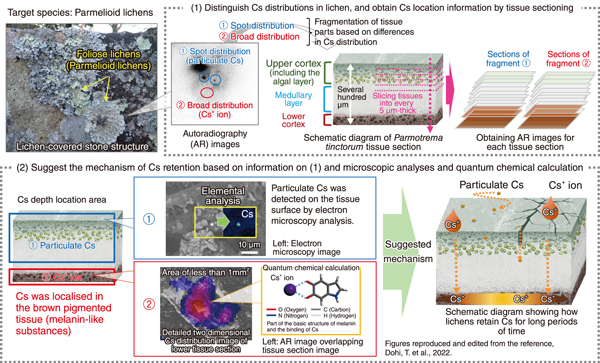
Fig.1 Conceptual diagram analyzing how radiocaesium is retained in lichen thallus tissues
Lichens are widespread on the terrestrial ecosystem and are known for their longevity of several decades. They can capture and retain substances from the atmosphere through dust, rain, etc. They have been used as biomonitors to study environmental pollution in Japan and abroad owing to these properties.
We developed a method to understand the characteristics and spatial distribution of radiocaesium (Cs) fallout due to TEPCO’s Fukushima Daiichi Nuclear Power Station accident by using lichens (Fig.1). Lichens have long been known to accumulate Cs; however, the questions of “where” and “in what chemical form” do lichens retain radiocaesium in vivo has been a long mystery.
In our previous research, quantum chemical calculations suggested that Cs in lichens may be stabilized via the complexation of Cs+ ions with their specific secondary metabolites. Our target species, parmelioid lichens, have a distinct layered structure with tissue thickness of several hundred micrometers and different metabolites in each layer. We were inspired by the idea that if the detailed Cs distribution in each tissue layer can be obtained, the metabolites contributing to Cs retention can be estimated.
First, autoradiography (AR) images visualizing the Cs distribution in lichens were obtained. The images were classified into “broad distribution” and “spot distribution” caused by Cs+ ions and particulate Cs, respectively, based on previous studies (Fig.1(1)). Second, each part was sliced into 5-μm-thick tissue sections and their AR images were obtained again. This slice process enabled understanding the depth distribution of Cs in the tissue at a spatial resolution of 5 μm, which is better than the usually attained resolution of 25 μm in AR. Third, detailed two-dimensional images of Cs distribution and pigmented tissues were successfully obtained by overlapping the sectioned AR images with the digital microscopy images containing information on tissue location and color. Thus, we found, for the first time, that the Cs+ ion is localized in the pigmented parts of the tissue, while particulate Cs is localized on/in the tissue (Fig.1(2)). We suggested retention mechanisms of Cs in lichens for long periods based on the Cs localization information, quantum chemical calculations, and electron microscopy analyses. Cs+ ions can be captured via complexation with melanin-like substances. Cs particles can be trapped on tissue surface via the development of hyphae and can be retained inside the tissue physically.
Using these methods, we can assume the chemical form of fallout Cs (deposited on lichens); thus, lichens act as a recorder of Cs.
This study is a part of a joint research project with the National Museum of Nature and Science. This research was supported by JSPS KAKENHI Grant-in-Aid for Challenging Exploratory Research (JP16K12627). Electron microscopy analyses were performed in cooperation with Hitachi High-Tech Corporation and JEOL Ltd.
(Terumi Dohi)

