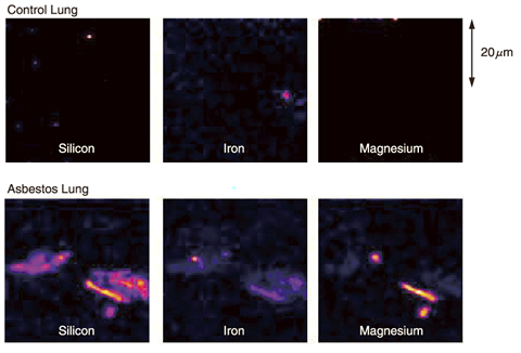
Fig.14-22 Pulmonary tissue taken from lung cancer patients with and w/o asbestos
Asbestos is called a "silent time bomb" because the asbestos-related symptoms appear after scores of years. Therefore, investigations of type, concentration and distribution of asbestos in lungs are very important for early recognition and treatment. However, in the present situation, surgical extraction of about 5g of lung tissue is necessary for diagnosis of asbestos-related lung diseases.
Particle induced X-ray emission (PIXE) is a powerful and nondestructive elemental analysis technique. Bombardment by ions with sufficient energy causes inner shell ionization of target atoms in a specimen. When the outer-shell-electrons drop down to the induced inner-shell vacancies, X-rays with energies unique to each element are emitted. By analyzing the energy spectrum of the X-rays, the elements in the specimen can be identified, and also the absolute concentration and distribution of each element can be measured. This method has about 100 times higher sensitivity than electron microprobe analysis (EPMA), because the electrons in the target atom are more efficiently removed by ions than by electrons. Combining these advantages of PIXE with our extremely thin (dia. less than 1μm) ion beam formation technique, a technique of in-air micro-PIXE analysis has been developed. This method can analyze distribution of about 80 elements from magnesium to uranium in a minute specimen of 100μm or less.
In this study, the position and shape of asbestos fibers in lung tissue were successfully identified through elemental analysis by in-air micro-PIXE. In addition, the type of asbestos was identified from the proportion of the elements (Fig.14-22).
This study was carried out in collaboration with Gunma University as part of its 21st century COE program "Biomedical Research Using Accelerator Technology". These results were published in an international medical journal. After this study, the correlation of the distributions of silicon, a main element of asbestos, and of the protein involved with pulmonary fibrosis was elucidated by means of in-air micro-PIXE combined with immunohistochemical staining.

