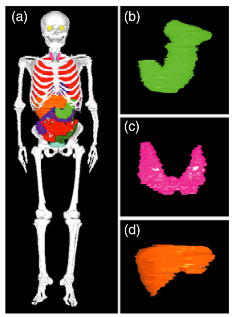
Fig.7-16 Three dimensional anterior views of (a) whole body, (b) stomach, (c) thyroid and (d) liver of Japanese voxel phantom in upright posture
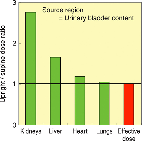
Fig.7-17 Comparison of organ doses and effective doses due to the intake of 131Cs when upright and when supine
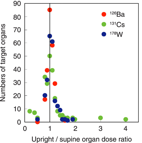
Fig.7-18 Distribution of the upright/supine organ dose ratios due to the intake of 126Ba, 131Cs and 178W
In order to estimate the health effects in humans due to radiation exposures, organ dose needs to be calculated using human models which reproduce body sizes and shapes and elemental compositions of organs. In recent years, voxel phantoms (phantoms), which consist of small rectangular block units called "voxel", have been developed on the basis of medical images of actual persons of various races, ages and body sizes, and are used for dose assessment. The reference phantoms adopted by the International Commission on Radiological Protection (ICRP) have been constructed on the basis of Caucasian anatomical data. The ICRP reference phantoms will be used to calculate organ doses for the purposes of radiation protection.
During radiation work, the posture is usually upright. However, the posture of previously developed phantoms is supine only. This is because the medical images are usually obtained from supine subjects. Therefore, it was impossible to clarify the variation in organ doses caused by changes in posture. We developed a Japanese adult male voxel phantom with upright posture on the basis of CT images of upright subjects for the first time in the world (Fig.7-16). The CT images in upright were again obtained from a subject for the phantom in supine previously developed by JAEA. Therefore, it is possible using these two phantoms to compare directly organ doses and to analyze the effects of posture on dose assessment. The voxel size of these phantoms is about 1mm3, which enables us to accurately represent e.g. the thyroid with its complicated shape.
Fig.7-17 shows examples of organ doses calculated using upright and supine phantoms. The upright/supine ratio of doses in the kidneys and liver were about 270% and 170%, respectively, larger than those of heart and lungs. This result was due to the differences in the movement distances of organs when changing posture; the movement distances of kidneys and liver due to the changes of posture were longer than those of heart and lungs. The organ doses were changed by the changes in posture, but differences in the organ doses are within a factor of 2 in most cases (Fig.7-18). These results indicate that the impact of posture on the organ doses is not significant. Therefore, it can be concluded that there is no problem in using ICRP reference phantoms which have been developed from tomographic images obtained in the supine posture for the calculation of dosimetric quantities in radiation protection. The knowledge obtained from this study was reflected in the ICRP Publication 110 dealing with adult reference computational phantoms.

