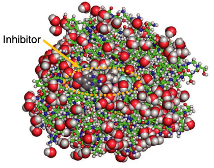
Fig.4-22 Whole atom positions in elastase complexed with inhibitor
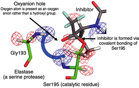
Fig.4-23 Oxyanion hole in the active center of elastase observed by neutron diffraction
Almost one third of all proteases can be classified as serine proteases, named for the nucleophilic serine residue at the active site. The tetrahedral intermediate in catalytic reactions is considered to stabilize electrostatically though hydrogen bonds with the backbone amides of Gly193 and Ser195, which together form an “oxyanion hole.” However, the state of the oxygen atom of the substrate in the oxyanion hole has not been confirmed yet. To help resolve long-standing questions regarding the catalytic activity of the serine proteases, the structure of porcine pancreatic elastase (PPE), which has been frequently used for research in structure-based drug design (SBDD), was analyzed by high-resolution neutron crystallography, because neutrons strongly interact with hydrogen and deuterium atoms and the neutron scattering lengths of hydrogen and deuterium atoms are very similar to those of other atoms.
We have succeeded in determining the positions of all the atoms of PPE complexed with its inhibitor by neutron crystallography at 1.65 Å resolution using the BIX-3 diffractometer installed at the 1G-A port of the research reactor JRR-3 (Fig.4-22). The inhibitor used in this study was covalently bound to the side chain of the Ser195 of the PPE. In this covalent complex, the carbonyl structure in the inhibitor was converted to a tetrahedral structure mimicking the catalytic transition intermediate state.
The carbonyl oxygen of the oxopropyl group of the inhibitor was converted to a hydroxyl oxygen that interacts with the oxyanion hole, comprising two hydrogen atoms from the backbone amides of Gly193 and Ser195. These two hydrogen atoms (deuterium in the neutron structure) were clearly confirmed in the Fo-Fc nuclear density maps (Fig.4-23). The nuclear density maps also show that the oxygen is present as an oxygen anion rather than a hydroxyl group, supporting the role of the oxyanion hole in stabilizing the tetrahedral intermediate in catalysis.
This is the first case in which an oxyanion of an intermediate analogue has been directly observed at an oxyanion hole. The precise structural information including hydrogen positions will contribute to further elucidation of the catalytic mechanism of serine protease. In addition, the structural information including hydrogen positions will provide additional knowledge to improve inhibitor design for therapeutic application with the fatal disease pancreatitis.

