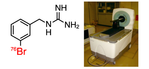
Fig.4-25 Chemical structure of 76Br-MBBG (left) and photo of a small-animal PET (right)
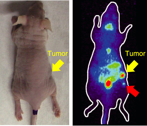
Fig.4-26 Photo of a pheochromocytoma-bearing mouse (left) and PET imaging
at 3 h after administration of 76Br-MBBG (right)
Pheochromocytoma is a tumor in the medulla of the adrenal glands. This tumor secretes excessive amounts of catecholamines such as epinephrine (adrenaline), which causes heavy hypertension. Although pheochromocytoma is usually curable by surgical resection, patients with small lesions or multiple metastases will be fatal because other treatments are not effective. Thus, early detection is critical to cure the pheochromocytoma. However, it is difficult to detect a small lesion or early metastasis using X-ray computed tomography (CT) or single photon emission computed tomography (SPECT) due to their lower spatial resolution.
We focused on positron emission tomography (PET) to overcome the above-mentioned problems. PET is a nuclear imaging technology that images distribution of lesions or metabolic activity in tissues of interest by detecting γ-rays from positron emitters. PET has a high potential to detect small tumors, since it has a higher spatial resolution compared to CT and SPECT. A positron emitter labeled compound that shows specific uptake into pheochromocytoma thus can be a promising tracer for detecting small pheochromocytoma using PET. We synthesized the positron emitter Br-76 (76Br) labeled 76Br-meta-bromobenzylguanidine (76Br-MBBG), which has high affinity to pheochromocytoma.
76Br-MBBG was administrated to tumor bearing mice and PET scans were performed at 3 h after administration (Fig.4-25). As a result, transplanted tumors were successfully imaged using 76Br-MBBG. Furthermore, a small tumor (size: 2 mm) undetected before PET scans was clearly imaged (Fig.4-26). These results indicated that 76Br-MBBG is a potential radiopharmaceutical for imaging pheochromocytoma and detecting very small tumors.
76Br-MBBG could be a powerful tool for early detection of pheochromocytoma. Furthermore, 76Br-MBBG could also be provided for imaging neruoblastoma, medullary thyroid carcinoma, and carcinoid which specifically accumulate 76Br-MBBG like pheochromocytoma.
<Previous: 4-12 | Next: 5 Photo-Medical Research Cooperation >

