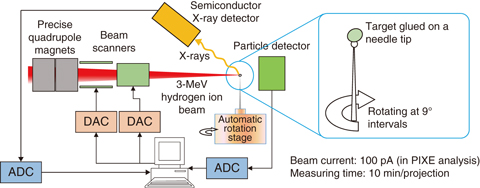
Fig.5-25 Schematic diagram of the PIXE Tomography system
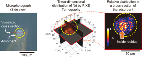
Fig.5-26 Optical micrograph of a porous silica-adsorbent particle for EXC (left figure) and the results of PIXE tomography (center and right figures)
We developed a particle-induced X-ray emission (PIXE) tomography system (Fig.5-25) based on the micro-PIXE analyzer using a MeV-class hydrogen-ion microbeam in the TIARA facility. In our system, a three-dimensional elemental distribution was obtained by image reconstruction from 40 two-dimensional projections measured by micro-PIXE. Moreover, to compensate for the decrease of X-ray production with decreasing energy of the beam and the absorption of X-rays, the modified maximum-likelihood-expectation-maximization (ML-EM) image-reconstruction method has enabled us to know the internal elemental distribution, even in a target several dozen μms thick.
Using our PIXE tomography system, we analyzed a 50-μm-diameter porous silica-adsorbent particle for extraction chromatography (EXC). In EXC, dissolved ions are first adsorbed to the adsorbent packed in a column and then eluted. Although EXC is promising for selectively removing long-half-life minor actinides (MAs) from high-level radioactive waste, its elution rate is insufficient at present. Even after the elution process, neodymium (Nd) substituting for MAs was detected by micro-PIXE. However, it was difficult to not only estimate the residual quantity inside the particle because of the overlapping surface distribution but also directly analyze a cross-sectional surface because of fragility. Therefore, we made an effort to measure the cross-sectional distribution of the residual Nd nondestructively using PIXE tomography. Consequently, we identified locally-concentrated Nd inside the eluted particle, as shown in Fig.5-26. Such a distribution is considered to depend upon the size of the micropores in the adsorbent, the concentration, and the passing speed of eluent. We will decide the optimal conditions of these parameters using PIXE tomography from now on.

